File:Arey1924 fig374.jpg

Original file (1,200 × 987 pixels, file size: 268 KB, MIME type: image/jpeg)
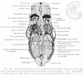
Fig. 374. Ventral reconstruction of the cardinal and subcardinal veins in a 6 mm pig embryo
Showing the early development of the inferior vena cava ( Vehe). X 22. In the small orientation figure (cf. Fig. 376) the various jdanes are indicated by broken lines.
The anterior cardinal veins (Figs. 374 and 375) are formed to drain the plexus of veins on each side of the head. These vessels extend caudad and lie ventro-lateral to the myelencephalon. Each receives branches from the sides of the myelencephalon, then curves ventrad, is joined by the linguo-facial vein from the branchial arches and at once unites with the posterior cardinal of the same side to form the common cardinal vein. This, as already explained, opens into the sinus venosus.
A posterior cardinal vein develops dorso-lateral to each mesonephros (Figs. 374 and 375). Running cephalad, they join the anterior cardinal veins. When the mesonephroi become prominent, as at this stage, the middle third of each posterior cardinal is broken up into sinusoids. Sinusoids extend from the posterior cardinal vein ventrally around both the lateral and medial surfaces of the mesonephros. The median sinusoids anastomose longitudinally and form the subcardinal veins, right and left.
| Historic Disclaimer - information about historic embryology pages |
|---|
| Pages where the terms "Historic" (textbooks, papers, people, recommendations) appear on this site, and sections within pages where this disclaimer appears, indicate that the content and scientific understanding are specific to the time of publication. This means that while some scientific descriptions are still accurate, the terminology and interpretation of the developmental mechanisms reflect the understanding at the time of original publication and those of the preceding periods, these terms, interpretations and recommendations may not reflect our current scientific understanding. (More? Embryology History | Historic Embryology Papers) |
Reference
Arey LB. Developmental Anatomy. (1924) W.B. Saunders Company, Philadelphia.
Cite this page: Hill, M.A. (2024, April 27) Embryology Arey1924 fig374.jpg. Retrieved from https://embryology.med.unsw.edu.au/embryology/index.php/File:Arey1924_fig374.jpg
- © Dr Mark Hill 2024, UNSW Embryology ISBN: 978 0 7334 2609 4 - UNSW CRICOS Provider Code No. 00098G
File history
Click on a date/time to view the file as it appeared at that time.
| Date/Time | Thumbnail | Dimensions | User | Comment | |
|---|---|---|---|---|---|
| current | 10:05, 23 October 2016 |  | 1,200 × 987 (268 KB) | Z8600021 (talk | contribs) | |
| 10:04, 23 October 2016 |  | 1,771 × 1,606 (554 KB) | Z8600021 (talk | contribs) | Fig. 374. Ventral reconstruction of the cardinal and subcardinal veins in a 6 mm. pig embryo, showing the early development of the inferior vena cava ( Vehe). X 22. In the small orientation figure (cf. Fig. 376) the various jdanes are indicated by brok... |
You cannot overwrite this file.
File usage
The following 2 pages use this file:

