File:Arey1924 fig229.jpg
From Embryology

Size of this preview: 800 × 500 pixels. Other resolution: 1,000 × 625 pixels.
Original file (1,000 × 625 pixels, file size: 150 KB, MIME type: image/jpeg)
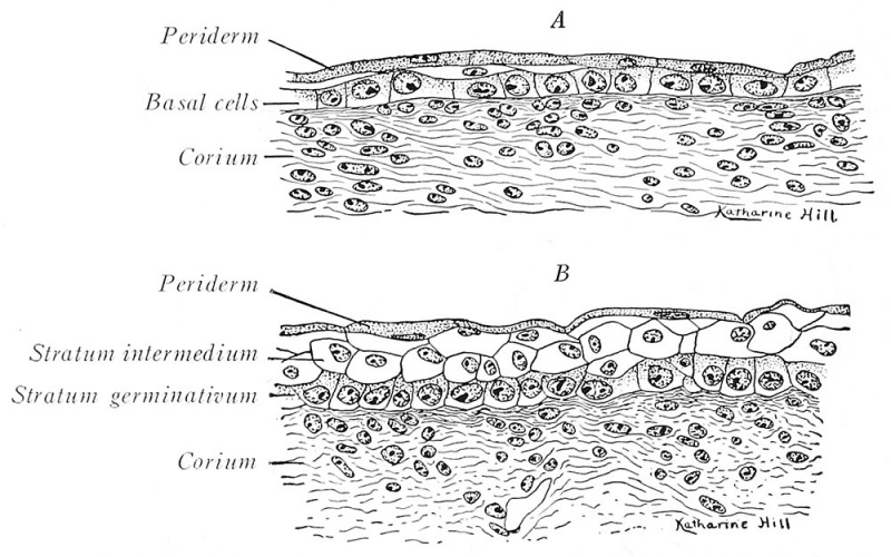
Fig. 229. Sections of the integument from a three-months fetus
(Prentiss) X 440
A. From the neck, showing at the right a two-layered epidermis and at tlie left the beginning of an intermediate layer;
B, from the chin, with three well-developed cjudermal layers.
| Historic Disclaimer - information about historic embryology pages |
|---|
 |

Reference
Arey LB. Developmental Anatomy. (1924) W.B. Saunders Company, Philadelphia.
Cite this page: Hill, M.A. (2024, April 27) Embryology Arey1924 fig229.jpg. Retrieved from https://embryology.med.unsw.edu.au/embryology/index.php/File:Arey1924_fig229.jpg
- © Dr Mark Hill 2024, UNSW Embryology ISBN: 978 0 7334 2609 4 - UNSW CRICOS Provider Code No. 00098G
File history
Click on a date/time to view the file as it appeared at that time.
| Date/Time | Thumbnail | Dimensions | User | Comment | |
|---|---|---|---|---|---|
| current | 12:40, 23 October 2016 | | 1,000 × 625 (150 KB) | Z8600021 (talk | contribs) | |
| 12:39, 23 October 2016 |  | 1,766 × 1,054 (317 KB) | Z8600021 (talk | contribs) | {{Arey1924 Footer}} Category:Integumentary |
You cannot overwrite this file.
File usage
The following 2 pages use this file: