File:Arey1924 fig202.jpg

Original file (1,200 × 599 pixels, file size: 153 KB, MIME type: image/jpeg)
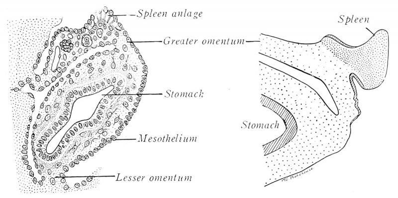
Fig. 202. Developmental stages of the human spleen
(redrawn from Kollman and Tonkoff). A, 10.5 mm.; B, 20 mm.
Embryos of 9 mm. exhibit a swelling on the left side of the dorsal mesogastrium, near the dorsal pancreas (Fig. 202 A). The thickening is due to a temporary proliferation and invasion of mesothelial cells into the underlying mesenchyme, which, meanwhile, has also undergone local enlargement and vascularization. These cells from the peritoneal epithelium give rise to a large part, at least, of the future spleen. The union of the splenic anlage with the mesogastrium (Fig. 202 B) is ultimately reduced to a narrow band.
At first the blood vessels constitute a closed system. The peculiar adult circulation is acquired relatively late. Lymphoid tissue first appears as ellipsoids about the smallest arteries in fetuses of four months. At seven months, the ovoid splenic corpuscles form nodules about the larger arteries. The capsule, trabeculce, and reticulum differentiate from the cells of the common anlage. During the last half of fetal life, red blood corpuscles are developed actively in the splenic capillaries.
| Historic Disclaimer - information about historic embryology pages |
|---|
 |

Reference
Arey LB. Developmental Anatomy. (1924) W.B. Saunders Company, Philadelphia.
Cite this page: Hill, M.A. (2024, April 27) Embryology Arey1924 fig202.jpg. Retrieved from https://embryology.med.unsw.edu.au/embryology/index.php/File:Arey1924_fig202.jpg
- © Dr Mark Hill 2024, UNSW Embryology ISBN: 978 0 7334 2609 4 - UNSW CRICOS Provider Code No. 00098G
File history
Click on a date/time to view the file as it appeared at that time.
| Date/Time | Thumbnail | Dimensions | User | Comment | |
|---|---|---|---|---|---|
| current | 13:49, 23 October 2016 | | 1,200 × 599 (153 KB) | Z8600021 (talk | contribs) | |
| 13:49, 23 October 2016 |  | 1,747 × 843 (231 KB) | Z8600021 (talk | contribs) | ==Fig. 202. Developmental stages of the human spleen== (redrawn from Kollman and Tonkoff). A, 10.5 mm.; B, 20 mm. {{Arey1924 Footer}} Category:Spleen |
You cannot overwrite this file.
File usage
The following 2 pages use this file: