File:Arey1924 fig195.jpg
From Embryology
Size of this preview: 800 × 480 pixels. Other resolution: 1,583 × 950 pixels.
Original file (1,583 × 950 pixels, file size: 267 KB, MIME type: image/jpeg)
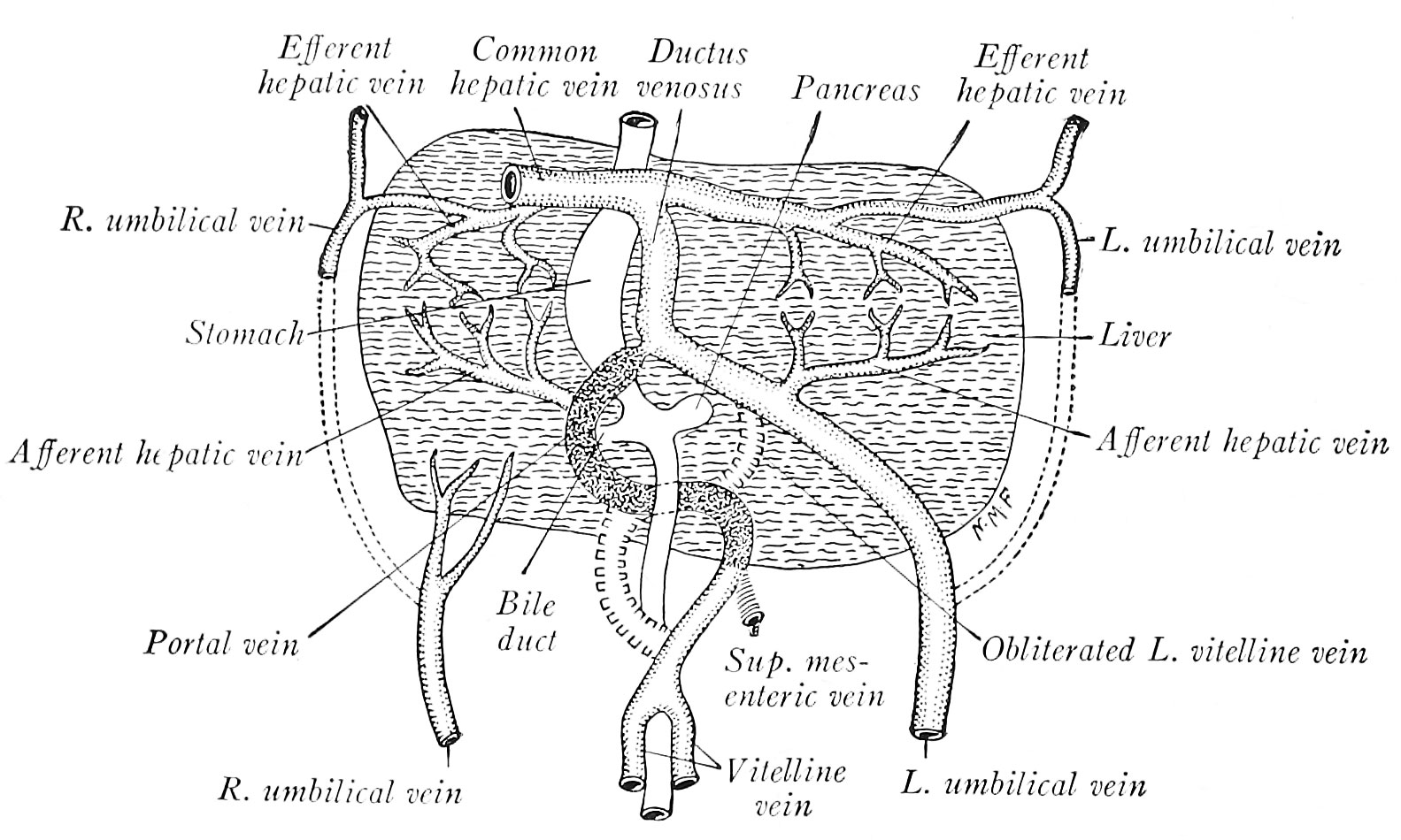
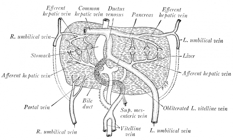
Fig. 195. The origin of the portal vein and ductus venosus as illustrated by a human embryo of 7 mm
(modified after His).
File history
Click on a date/time to view the file as it appeared at that time.
| Date/Time | Thumbnail | Dimensions | User | Comment | |
|---|---|---|---|---|---|
| current | 14:04, 23 October 2016 | | 1,583 × 950 (267 KB) | Z8600021 (talk | contribs) | |
| 14:02, 23 October 2016 | 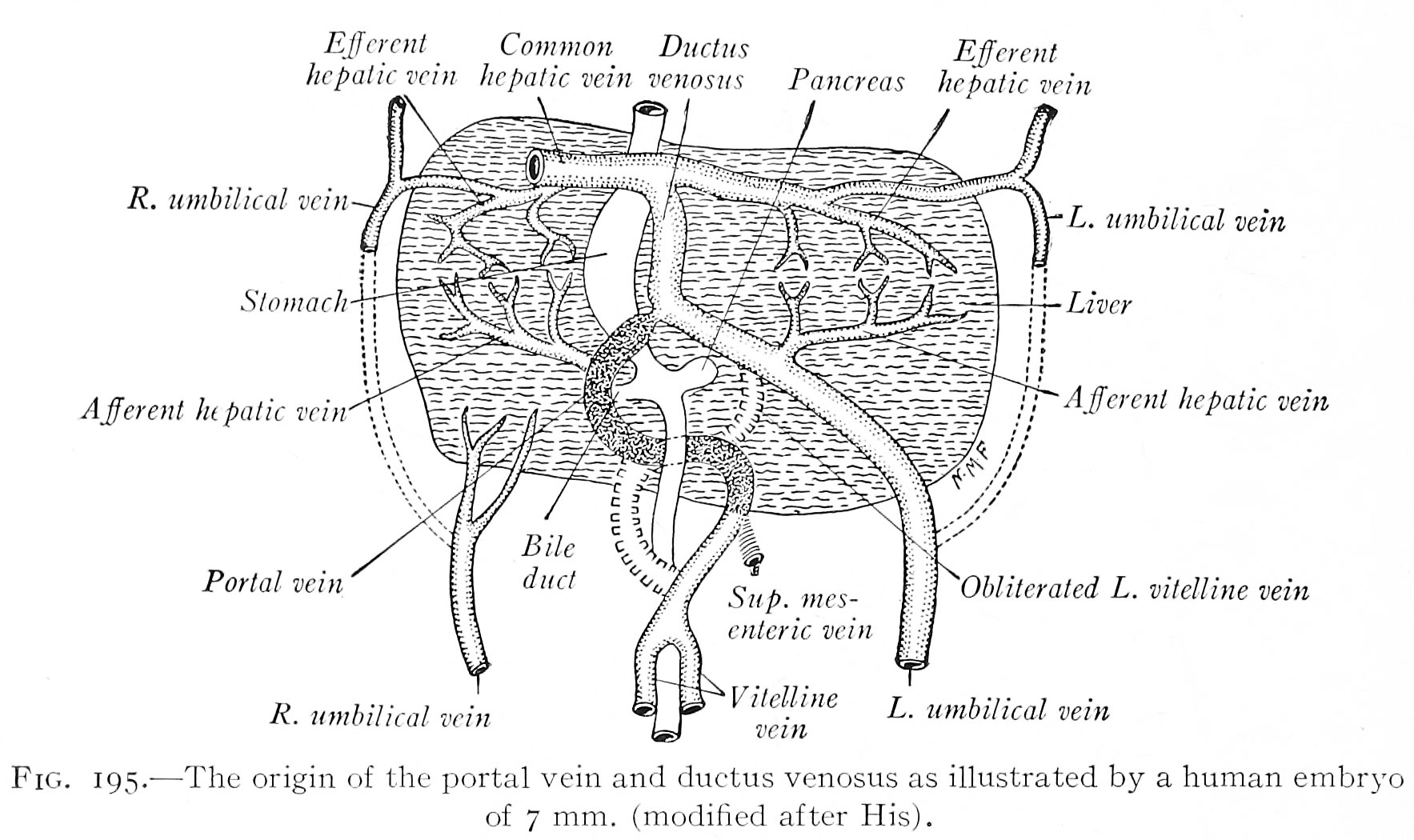 | 1,767 × 1,050 (297 KB) | Z8600021 (talk | contribs) | ==Fig. 195. The origin of the portal vein and ductus venosus as illustrated by a human embryo of 7 mm== (modified after His). |
You cannot overwrite this file.
File usage
The following 2 pages use this file: