File:Arey1924 fig010.jpg
From Embryology

Size of this preview: 486 × 599 pixels. Other resolution: 852 × 1,050 pixels.
Original file (852 × 1,050 pixels, file size: 143 KB, MIME type: image/jpeg)
Fig. 10. Diagrams of the development of spermatozoa
(Meves in Lewis and Stohr). a.c, Anterior centrosome; a.f., axial filament; c.p., connecting piece; ch.p., chief piece; g.c., cap; n., nucleus; nk., neck; p., cytoplasm; p.c., posterior centrosome.
| Historic Disclaimer - information about historic embryology pages |
|---|
 |

Reference
Arey LB. Developmental Anatomy. (1924) W.B. Saunders Company, Philadelphia.
Cite this page: Hill, M.A. (2024, May 19) Embryology Arey1924 fig010.jpg. Retrieved from https://embryology.med.unsw.edu.au/embryology/index.php/File:Arey1924_fig010.jpg
- © Dr Mark Hill 2024, UNSW Embryology ISBN: 978 0 7334 2609 4 - UNSW CRICOS Provider Code No. 00098G
File history
Click on a date/time to view the file as it appeared at that time.
| Date/Time | Thumbnail | Dimensions | User | Comment | |
|---|---|---|---|---|---|
| current | 17:26, 4 January 2017 | | 852 × 1,050 (143 KB) | Z8600021 (talk | contribs) | |
| 17:25, 4 January 2017 | 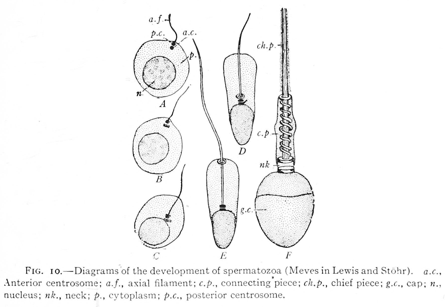 | 1,774 × 1,227 (209 KB) | Z8600021 (talk | contribs) |
You cannot overwrite this file.
File usage
The following 2 pages use this file: