File:Aorta coarctation echocardiogram.jpg: Difference between revisions
mNo edit summary |
mNo edit summary |
||
| Line 6: | Line 6: | ||
'''B''' Colour Doppler of the same image with aliasing of flow at the site of coarctation (arrow). | '''B''' Colour Doppler of the same image with aliasing of flow at the site of coarctation (arrow). | ||
:'''Links:''' {{coarctation of the aorta}} | |||
===Reference=== | ===Reference=== | ||
Latest revision as of 14:55, 8 March 2019
Coarctation of the Aorta Echocardiogram
Echocardiography uses standard two-dimensional, three-dimensional, and Doppler ultrasound to create images of the heart.
A Two-dimensional transthoracic echocardiogram image obtained from the suprasternal notch in an 11-day-old infant demonstrating discrete coarctation (arrow);
B Colour Doppler of the same image with aliasing of flow at the site of coarctation (arrow).
- Links: coarctation of the aorta
Reference
Torok RD, Campbell MJ, Fleming GA & Hill KD. (2015). Coarctation of the aorta: Management from infancy to adulthood. World J Cardiol , 7, 765-75. PMID: 26635924 DOI.
Copyright
Open-Access: This article is an open-access article which was selected by an in-house editor and fully peer-reviewed by external reviewers. It is distributed in accordance with the Creative Commons Attribution Non Commercial (CC BY-NC 4.0) license, which permits others to distribute, remix, adapt, build upon this work non-commercially, and license their derivative works on different terms, provided the original work is properly cited and the use is non-commercial. See: http://creativecommons.org/licenses/by-nc/4.0/
Figure 1 WJC-7-765-g001.jpg
Cite this page: Hill, M.A. (2024, May 21) Embryology Aorta coarctation echocardiogram.jpg. Retrieved from https://embryology.med.unsw.edu.au/embryology/index.php/File:Aorta_coarctation_echocardiogram.jpg
- © Dr Mark Hill 2024, UNSW Embryology ISBN: 978 0 7334 2609 4 - UNSW CRICOS Provider Code No. 00098G
File history
Click on a date/time to view the file as it appeared at that time.
| Date/Time | Thumbnail | Dimensions | User | Comment | |
|---|---|---|---|---|---|
| current | 14:40, 8 March 2019 | 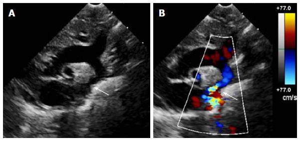 | 601 × 283 (28 KB) | Z8600021 (talk | contribs) | ==Echocardiogram of coarctation== A: Two-dimensional transthoracic echocardiogram image obtained from the suprasternal notch in an 11-day-old infant demonstrating discrete coarctation (arrow); B: Colour Doppler of the same image with aliasing of flo... |
You cannot overwrite this file.
File usage
The following page uses this file: