File:AnsonKarabinMartin1939 fig60-64.jpg
From Embryology

Size of this preview: 513 × 600 pixels. Other resolution: 1,280 × 1,496 pixels.
Original file (1,280 × 1,496 pixels, file size: 130 KB, MIME type: image/jpeg)
Figs. 60 to 64. Reconstructions of the fissula ante fenestram; anterolateral views
X 17. figure 60, embryo of 135 mm.; figure 61, embryo of 161 mm.: figure 62, embryo of 183 mm.; figure 63, infant 3 months old, and figure 64, adult 70 years old.
The unlabeled arrows point to the tympanic orifices of the fissulae; the additional, lower, arrow in figure 64 marks the point of junction of cartilage and fibrous tissue.
Reference
Anson BJ. Karabin JE. and Martin J. Stapes, fissula ante fenestram and associated structures in man: II. From Fetus at Term to Adult of Seventy (1938) Arch. Otolaryng. 28: 676-697.
File history
Click on a date/time to view the file as it appeared at that time.
| Date/Time | Thumbnail | Dimensions | User | Comment | |
|---|---|---|---|---|---|
| current | 10:01, 18 October 2017 | | 1,280 × 1,496 (130 KB) | Z8600021 (talk | contribs) | |
| 09:55, 18 October 2017 | 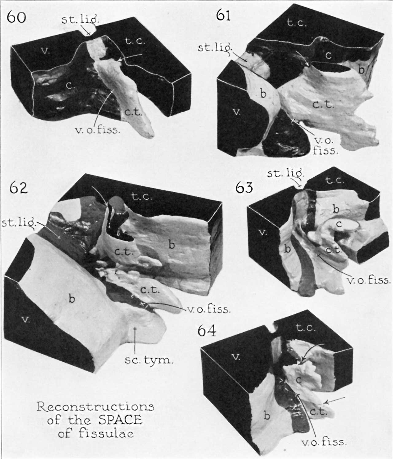 | 1,280 × 1,496 (136 KB) | Z8600021 (talk | contribs) | ||
| 09:54, 18 October 2017 |  | 1,635 × 2,187 (459 KB) | Z8600021 (talk | contribs) | ===Reference=== {{Ref-AnsonKarabinMartin1939}} |
You cannot overwrite this file.
File usage
The following page uses this file: