File:Anson1948 fig14.jpg

Original file (1,280 × 872 pixels, file size: 190 KB, MIME type: image/jpeg)
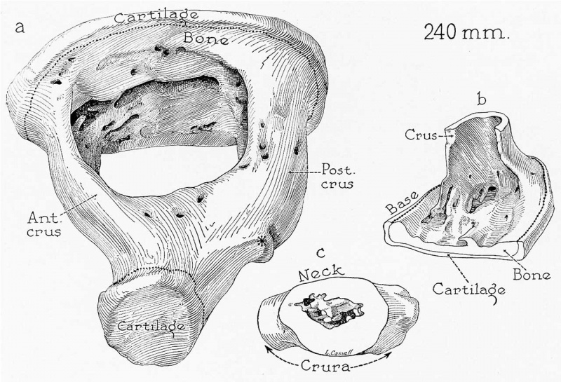
Fig. 14. Drawings of a reconstruction of the stapes in a 240 mm
(27 week) fetus (Wisconsin series 112); x 7: (a) reconstruction pictured entire and viewed from a superolateral position; (b) the basal portion of the anterior crus and the adjacent anterior third of the base seen as though from the obturator space; (c) the neck of the stapes (capital portion removed) and the adjacent portions of the two crura, in lateral view. In a are shown the limits of the portions shown in b and c.
The crura are now channeled columns (a and b) and are foraminous in certain areas (a). On the base of the ossicle some of the persistent perichondrial bone has formed an incomplete crista stapedis (a and 12); under arrow in (c). The neck and head are hollowed; yet some endochondral bone, in the form of spicules, remains to close the capital space (c). The stapes has assumed typical adult form: The anterior crus is the shorter and thinner of the two; a pronounced tubercle appears on the posterior crus, for attachment of the tendon of the stapedius muscle, and the head is foveate on its articular surface (at). The osseous lamina of the base is composed of endochondral bone, with which have fused remnants of the original perichondrial bone from the once complete obturator wall of the base (a and b). As is regularly the case, the developmental changes affecting the head of the stapes are retarded; part of the obturator wall here remains to close the capital space medially (c). Such bone sometimes persists in the stapes of adults, either in the form of a perforate plate or in that of osseous bridges.
Reference
Anson BJ. and Cauldwell EW. Stapes, fissula ante fenestram and associated structures in man: V . From the fetus of 160 mm to term. (1948) 48(3): 263-300.
Cite this page: Hill, M.A. (2024, April 26) Embryology Anson1948 fig14.jpg. Retrieved from https://embryology.med.unsw.edu.au/embryology/index.php/File:Anson1948_fig14.jpg
- © Dr Mark Hill 2024, UNSW Embryology ISBN: 978 0 7334 2609 4 - UNSW CRICOS Provider Code No. 00098G
File history
Click on a date/time to view the file as it appeared at that time.
| Date/Time | Thumbnail | Dimensions | User | Comment | |
|---|---|---|---|---|---|
| current | 21:28, 16 October 2017 | | 1,280 × 872 (190 KB) | Z8600021 (talk | contribs) | |
| 21:27, 16 October 2017 | 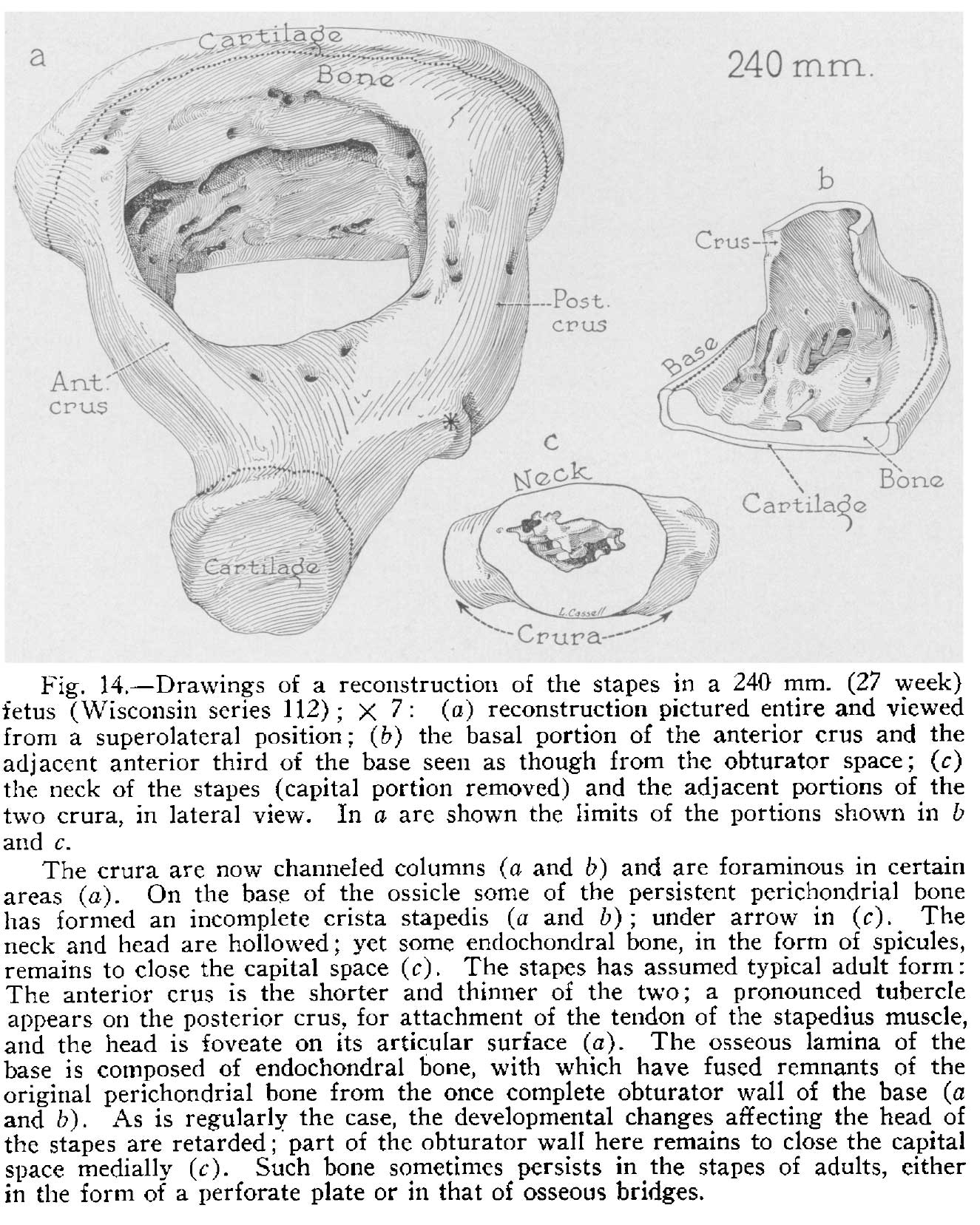 | 1,325 × 1,657 (358 KB) | Z8600021 (talk | contribs) |
You cannot overwrite this file.
File usage
The following 2 pages use this file: