File:Anson1948 fig09.jpg

Original file (1,280 × 1,686 pixels, file size: 235 KB, MIME type: image/jpeg)
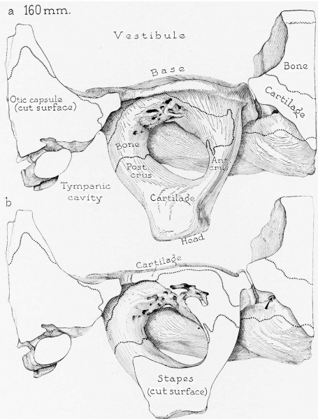
Fig. 9. Otic capsule developmental stages 160 mm fetus
Drawings of a reconstruction of the stapes and adjacent portions of the otic capsule in a 160 mm. (19.5 week) fetus (Wisconsin series 41); superior (cranial) views; X 7: (a) of the stapes entire; (b) with an upper segment of the stapes removed (in the plane of the constituent transverse section), showing erosion of the stapedial base and of the basal extremities of the crura. The dotted lines on the stapes indicates the approximate limits of the ossifying area; to either side of the area bounded by these lines, the cartilage of the base (on the vestibular surface) and of the head, neck and capital extremities of the crura (toward the tympanic aspect) is as yet unaltered. Similarly, osseous and cartilaginous portions of the capsule are indicated. In I) the arrow enters the tympanic orifice of the fissula; in figure 10b the arrow enters the junction of the vestibular extremity and the body of the fissula.
Reference
Anson BJ. and Cauldwell EW. Stapes, fissula ante fenestram and associated structures in man: V . From the fetus of 160 mm to term. (1948) 48(3): 263-300.
Cite this page: Hill, M.A. (2024, April 26) Embryology Anson1948 fig09.jpg. Retrieved from https://embryology.med.unsw.edu.au/embryology/index.php/File:Anson1948_fig09.jpg
- © Dr Mark Hill 2024, UNSW Embryology ISBN: 978 0 7334 2609 4 - UNSW CRICOS Provider Code No. 00098G
File history
Click on a date/time to view the file as it appeared at that time.
| Date/Time | Thumbnail | Dimensions | User | Comment | |
|---|---|---|---|---|---|
| current | 20:37, 16 October 2017 | | 1,280 × 1,686 (235 KB) | Z8600021 (talk | contribs) | |
| 20:36, 16 October 2017 |  | 1,320 × 2,194 (352 KB) | Z8600021 (talk | contribs) |
You cannot overwrite this file.
File usage
The following 2 pages use this file: