File:Anson1948 fig07.jpg
Original file (1,280 × 1,801 pixels, file size: 219 KB, MIME type: image/jpeg)
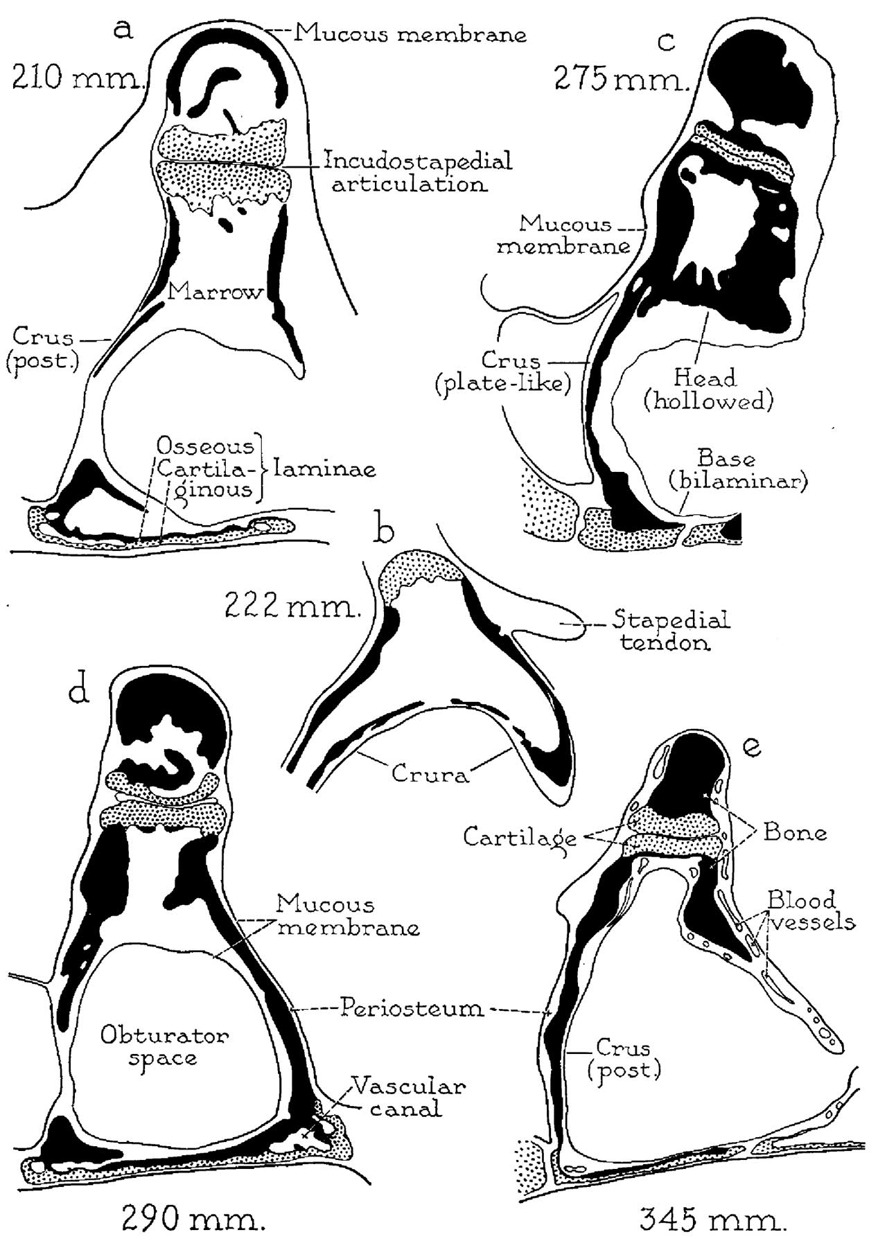
Fig. 7. Otic capsule developmental stages 210, 222, 275, 290 and 345 mm Fetus
Drawings of developmental stages, continued; X 6.6: (a) 210 mm. (23 week) fetus (Wisconsin series 51, slide 38, section 5); (b) 222 mm. (25 week) fetus (Wisconsin series 46, slide 19, section 10); (c) 275 mm. (30 week) fetus (Wisconsin series 4, slide 25, section 4); (d) 290 mm. (32 week) fetus (Vlfisconsin series 59, slide 36, section 5); (e) 345 mm. (38 week) fetus (Wisconsin series 61,. slide 42, section 1).
In the 275 mm specimen investment of the cartilaginous lamina of the stapedial base by endochondral bone has been completed (a) ; fusion of the peripheral remnant of perichondrial bone on the obturator aspect of the base (a) with the newly formed endochondral bone has resulted in the formation of osseous canals for the transmission of blood vessels (d). The mucous membrane and associated submucosal tissue, which in the 210 mm. specimen have already replaced the primitive marrow of the crura (a), later spread medialward to invest the endochondral and other bone of the base (290 mm., d); they ultimately invade the excavated neck and head of the stapes (345 mm., e). Thus, with regard to form, the stapes is essentially an adult ossicle in the 290 mm. fetus (d) ; with respect to mucosal relations, the ossicle attains adulthood at the 345 mm. stage (e) (compare with fig. 8e, from the newborn).
Reference
Anson BJ. and Cauldwell EW. Stapes, fissula ante fenestram and associated structures in man: V . From the fetus of 160 mm to term. (1948) 48(3): 263-300.
Cite this page: Hill, M.A. (2024, April 26) Embryology Anson1948 fig07.jpg. Retrieved from https://embryology.med.unsw.edu.au/embryology/index.php/File:Anson1948_fig07.jpg
- © Dr Mark Hill 2024, UNSW Embryology ISBN: 978 0 7334 2609 4 - UNSW CRICOS Provider Code No. 00098G
File history
Click on a date/time to view the file as it appeared at that time.
| Date/Time | Thumbnail | Dimensions | User | Comment | |
|---|---|---|---|---|---|
| current | 20:17, 16 October 2017 | | 1,280 × 1,801 (219 KB) | Z8600021 (talk | contribs) | |
| 20:09, 16 October 2017 |  | 1,481 × 2,211 (341 KB) | Z8600021 (talk | contribs) | ===Reference=== {{Ref-Anson1948}} {{Footer}} Category:Middle EarCategory:Historic EmbryologyCategory:1940's |
You cannot overwrite this file.
File usage
The following 2 pages use this file: