File:Anson1948 fig06.jpg
Original file (1,028 × 1,488 pixels, file size: 224 KB, MIME type: image/jpeg)
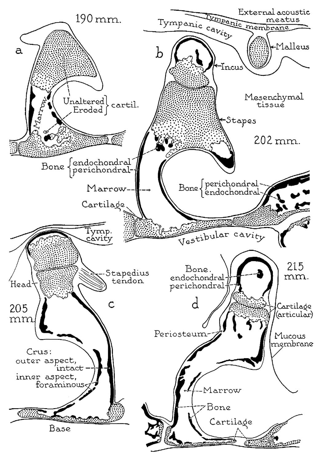
Fig. 6. Otic capsule developmental stages 190 mm and 215 mm fetus
Drawings of crucial developmental stages, continued: X 6.6. (a) 190 mm. (21 week) fetus (Wisconsin series 29A, slide 18, section 1); (b) 202 mm. (23 week) fetus (Wisconsin series 70, slide 27, section 6); (c) 205 mm. (23 week) fetus (Wisconsin series 129, slide 20, section 3); (d) 215 mm. (24 week) fetus (Wisconsin series 62, slide 28, section 4). Parts (1, b and d represent sections from series of the left ear; c is from the right ear; all represent the transverse level of the posterior crus of the stapes.
Four further steps in the progress of stapedial ossification are illustrated. In the 190 mm. specimen, the process of ossification has spread to the base of the stapes, but has not yet affected either the neck or the head of the ossicle (a). At ‘the 202 mm. stage excavation of the neck is in progress. (b) This developmental phase has been completed in the 205 mm. specimen; additionally, endochondral bone is being deposited on the internal surface of the excavated basal plate of cartilage. (c) In the 215 mm. fetus the articular plate of cartilage on the head of the stapes, now fully excavated, is being converted by a similar process into a bilaminar articulation. (d) Concurrently, a like series of changes is taking place in the lenticular process of the incus. Destruction of periosteal bone on the obturator surface of the stapes keeps pace with the formation of endochondral bone within the capital and basal portions of the ossicle.
Reference
Anson BJ. and Cauldwell EW. Stapes, fissula ante fenestram and associated structures in man: V . From the fetus of 160 mm to term. (1948) 48(3): 263-300.
Cite this page: Hill, M.A. (2024, April 26) Embryology Anson1948 fig06.jpg. Retrieved from https://embryology.med.unsw.edu.au/embryology/index.php/File:Anson1948_fig06.jpg
- © Dr Mark Hill 2024, UNSW Embryology ISBN: 978 0 7334 2609 4 - UNSW CRICOS Provider Code No. 00098G
File history
Click on a date/time to view the file as it appeared at that time.
| Date/Time | Thumbnail | Dimensions | User | Comment | |
|---|---|---|---|---|---|
| current | 20:04, 16 October 2017 | | 1,028 × 1,488 (224 KB) | Z8600021 (talk | contribs) | |
| 20:01, 16 October 2017 | 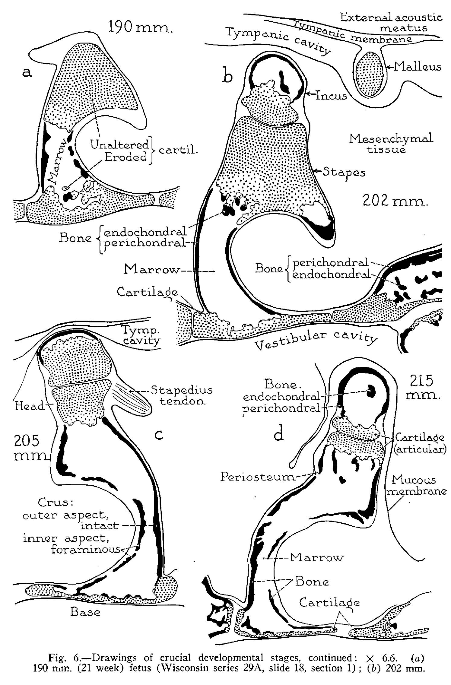 | 1,495 × 2,271 (470 KB) | Z8600021 (talk | contribs) | ===Reference=== {{Ref-Anson1948}} {{Footer}} Category:Middle EarCategory:Historic EmbryologyCategory:1940's |
You cannot overwrite this file.
File usage
The following 2 pages use this file: