File:Anson1948 fig04.jpg
Original file (1,280 × 1,541 pixels, file size: 247 KB, MIME type: image/jpeg)
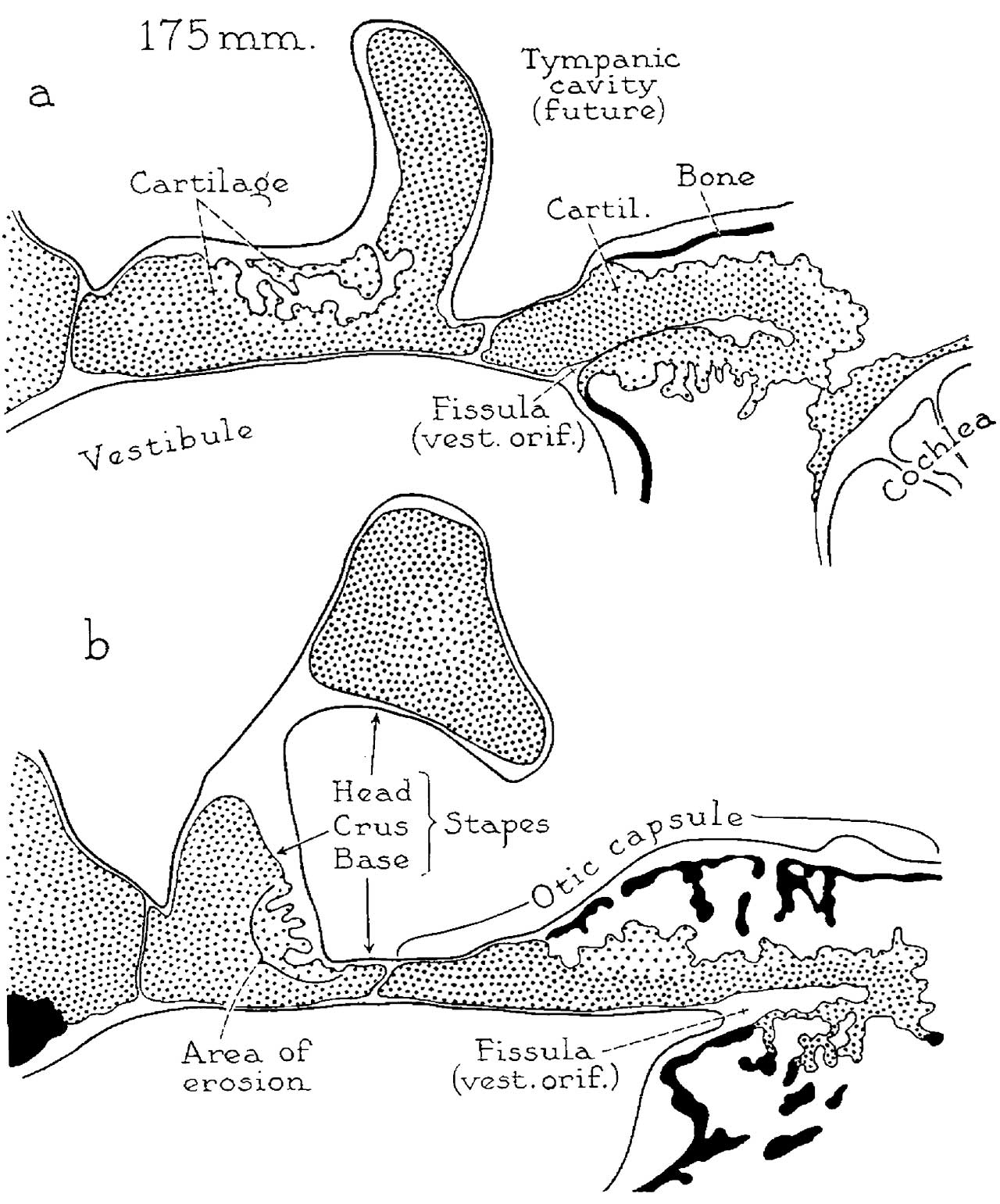
Fig. 4. Developmental stages 175 mm (20 week) fetus
Drawings (continued) of developmental stages from a 175 mm (20 week) fetus (Wisconsin series 104: (a) slide 17, section 5; (b) slide 16, section 2); x 6.6.
The stapes is now excavated on the obturator surface of the base (a and b), at the basal end of the anterior crus (a) and in the corresponding portion of the posterior crus (b). However, bone has not yet formed over the eroded area. In the antefenestral portion of the capsule, periosteal bone, which appears in the form of thin larninas, does not extend to the vestibular window. In the latter portion of the capsule the fenestral shell of cartilage is continuous with that which encloses the fissular tract of connective tissue.
The fissular shell of cartilage is becoming detached, at its opposite (or anterior) extremity, from the cartilaginous wall of the cochlea (a). In the region between the vestibule and the cochlea, spicules of intrachondrial bone now are present, having been formed as a result of rapid ossification of persistent spicules of cartilage (b).
Reference
Anson BJ. and Cauldwell EW. Stapes, fissula ante fenestram and associated structures in man: V . From the fetus of 160 mm to term. (1948) 48(3): 263-300.
Cite this page: Hill, M.A. (2024, April 26) Embryology Anson1948 fig04.jpg. Retrieved from https://embryology.med.unsw.edu.au/embryology/index.php/File:Anson1948_fig04.jpg
- © Dr Mark Hill 2024, UNSW Embryology ISBN: 978 0 7334 2609 4 - UNSW CRICOS Provider Code No. 00098G
File history
Click on a date/time to view the file as it appeared at that time.
| Date/Time | Thumbnail | Dimensions | User | Comment | |
|---|---|---|---|---|---|
| current | 09:20, 15 October 2017 | | 1,280 × 1,541 (247 KB) | Z8600021 (talk | contribs) | |
| 09:19, 15 October 2017 | 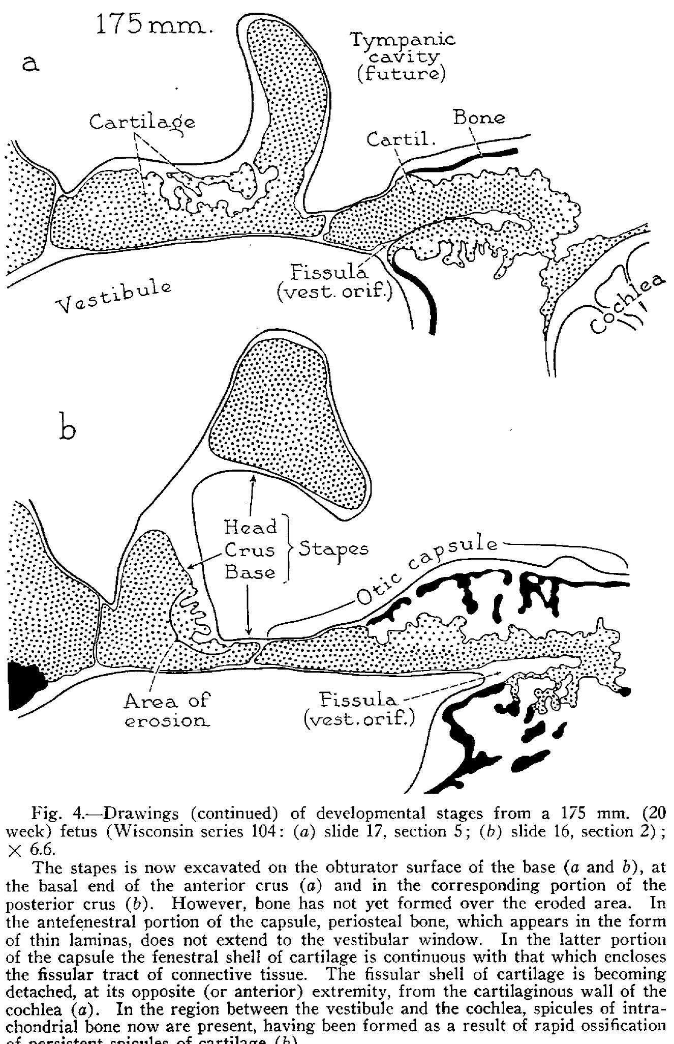 | 1,326 × 2,052 (444 KB) | Z8600021 (talk | contribs) |
You cannot overwrite this file.
File usage
The following 2 pages use this file: