File:Anson1934 fig19.jpg: Difference between revisions
(==Plate 2. Auditory Vesicle Models from Harvard Embryological Collection== Auditory vesicle and endolymphatic appendage in human embryos Figures 15 and 16, models prepared froiii a transverse series; figures 17, 18, and 19, from frontal series in the...) |
mNo edit summary |
||
| Line 1: | Line 1: | ||
== | ==Fig. 19. Auditory Vesicle Model from Harvard Embryological Collection== | ||
Auditory vesicle and endolymphatic appendage in human embryos Figures 15 and 16, models prepared froiii a transverse series; figures 17, 18, and 19, from frontal series in the Harvard Embryological Collection. Figures 15 and 16, X 130; 17, 18, and 19, X 45. | Auditory vesicle and endolymphatic appendage in human embryos Figures 15 and 16, models prepared froiii a transverse series; figures 17, 18, and 19, from frontal series in the Harvard Embryological Collection. Figures 15 and 16, X 130; 17, 18, and 19, X 45. | ||
* '''15''' - 8 mm., 2066. The endolymphatic appendage has lengtliened, and is now pyramidal in outline. It no longer bears any remnant of the stalk by which the otocyst arose. | * [[:File:Anson1934 fig15.jpg|'''15''']] - 8 mm., 2066. The endolymphatic appendage has lengtliened, and is now pyramidal in outline. It no longer bears any remnant of the stalk by which the otocyst arose. | ||
* '''16''' - The same model viewed from the front. Note the demarcating crease at the arrow, and the atiscvice of a nodule on the endolymphatic appendage. | * [[:File:Anson1934 fig16.jpg|'''16''']] - The same model viewed from the front. Note the demarcating crease at the arrow, and the atiscvice of a nodule on the endolymphatic appendage. | ||
* '''17''' - 12 mm., 816. Frontal series. Model, viewed froin behind; cut surface black. Compare the 8-mm. stage. At 12 mm., the appendage is not only considerably lengthened, but the two primary portions, the enlarged saccus and narrower ductus, are readily distiiiguisliablc from each other; these together are marked off sharply from the utricle by a crease (at the arrow) which constitutes the utricular fold or ‘valve.’ | * [[:File:Anson1934 fig17.jpg|'''17''']] - 12 mm., 816. Frontal series. Model, viewed froin behind; cut surface black. Compare the 8-mm. stage. At 12 mm., the appendage is not only considerably lengthened, but the two primary portions, the enlarged saccus and narrower ductus, are readily distiiiguisliablc from each other; these together are marked off sharply from the utricle by a crease (at the arrow) which constitutes the utricular fold or ‘valve.’ | ||
* '''18 and 19''' - Models from the 29-mm. and tlie 40-mnn. human embryos (frontal series, 914 and 1917 respectively). In the model of the 40-mm. embryo the proximal two-thirds of the endolymphatic duct is omitted. Lateral views. The view distal projection of the endolymphatic sac is well defined; this appendage is a separate growth, and not a derivative of the original stalk of connection between the vesicle and the ectoderm. | * [[:File:Anson1934 fig18.jpg|'''18''']] and [[:File:Anson1934 fig19.jpg|'''19''']] - Models from the 29-mm. and tlie 40-mnn. human embryos (frontal series, 914 and 1917 respectively). In the model of the 40-mm. embryo the proximal two-thirds of the endolymphatic duct is omitted. Lateral views. The view distal projection of the endolymphatic sac is well defined; this appendage is a separate growth, and not a derivative of the original stalk of connection between the vesicle and the ectoderm. | ||
{{Anson1934 figures}} | {{Anson1934 figures}} | ||
Revision as of 15:41, 23 July 2015
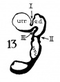
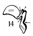
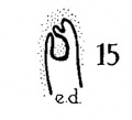
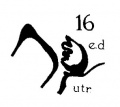
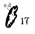
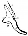
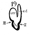
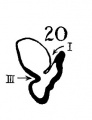
Fig. 19. Auditory Vesicle Model from Harvard Embryological Collection
Auditory vesicle and endolymphatic appendage in human embryos Figures 15 and 16, models prepared froiii a transverse series; figures 17, 18, and 19, from frontal series in the Harvard Embryological Collection. Figures 15 and 16, X 130; 17, 18, and 19, X 45.
- 15 - 8 mm., 2066. The endolymphatic appendage has lengtliened, and is now pyramidal in outline. It no longer bears any remnant of the stalk by which the otocyst arose.
- 16 - The same model viewed from the front. Note the demarcating crease at the arrow, and the atiscvice of a nodule on the endolymphatic appendage.
- 17 - 12 mm., 816. Frontal series. Model, viewed froin behind; cut surface black. Compare the 8-mm. stage. At 12 mm., the appendage is not only considerably lengthened, but the two primary portions, the enlarged saccus and narrower ductus, are readily distiiiguisliablc from each other; these together are marked off sharply from the utricle by a crease (at the arrow) which constitutes the utricular fold or ‘valve.’
- 18 and 19 - Models from the 29-mm. and tlie 40-mnn. human embryos (frontal series, 914 and 1917 respectively). In the model of the 40-mm. embryo the proximal two-thirds of the endolymphatic duct is omitted. Lateral views. The view distal projection of the endolymphatic sac is well defined; this appendage is a separate growth, and not a derivative of the original stalk of connection between the vesicle and the ectoderm.
| Historic Disclaimer - information about historic embryology pages |
|---|
 |
- Links: fig 1-7 | fig 1 | fig 2 | fig 3 | fig 4 | fig 5 | fig 6 | fig 7 | fig 8-21 | fig 22-33 | 1934 Anson | Historic Papers | Inner Ear Development
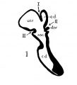
fig 1 Human 22.8 mm
fig 2 Human 29 mm
fig 3 Human 34 mm
fig 4 Human 40 mm
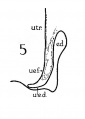
fig 5 Infant 4 months
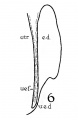
fig 6 Child 3 years
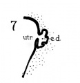
fig 7 embryo 36 mm
8 Cat 7 mm
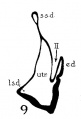
9 Cat 10.6 mm
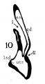
10 Cat 14 mm
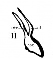
11 Cat 15 mm
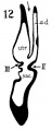
12 Cat 24.1 mm
13 Cat 32.6 mm
14 Cat 39 mm
15 Cat 39 mm
16 Cat 31 mm
17 Guinea-pig 18.5 mm
18 Dog 12.5 mm
19 Rabbit 21 mm
20 Rabbit 25 mm
21 Rabbit 29 mm


Reference
Anson BJ. The early development of the membranous labyrinth in mammalian embryos, with special reference to the endolymphatic duct and the utriculo—endolymphatic duct. (1934) Anat. Rec. 59: 15-25.
Cite this page: Hill, M.A. (2024, May 20) Embryology Anson1934 fig19.jpg. Retrieved from https://embryology.med.unsw.edu.au/embryology/index.php/File:Anson1934_fig19.jpg
- © Dr Mark Hill 2024, UNSW Embryology ISBN: 978 0 7334 2609 4 - UNSW CRICOS Provider Code No. 00098G
File history
Click on a date/time to view the file as it appeared at that time.
| Date/Time | Thumbnail | Dimensions | User | Comment | |
|---|---|---|---|---|---|
| current | 15:38, 23 July 2015 | 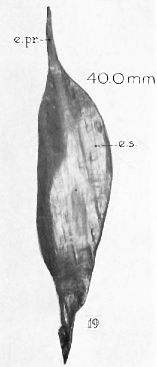 | 524 × 1,218 (48 KB) | Z8600021 (talk | contribs) | ==Plate 2. Auditory Vesicle Models from Harvard Embryological Collection== Auditory vesicle and endolymphatic appendage in human embryos Figures 15 and 16, models prepared froiii a transverse series; figures 17, 18, and 19, from frontal series in the... |
You cannot overwrite this file.
File usage
The following 3 pages use this file: