File:Aasar1931 plate3.jpg
Original file (1,507 × 2,284 pixels, file size: 680 KB, MIME type: image/jpeg)
Plate 3
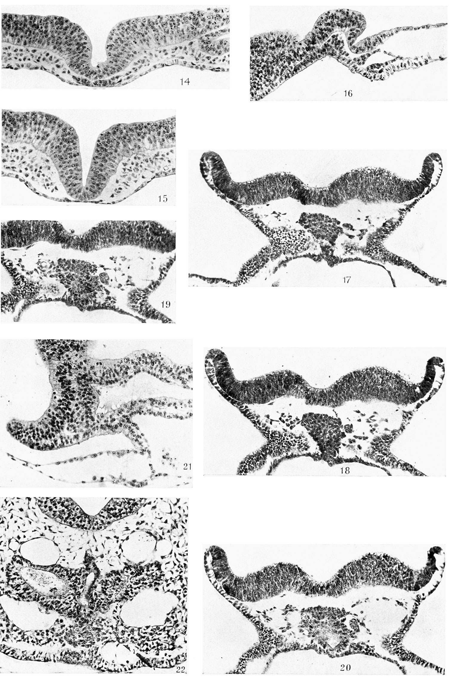
Fig. 14. 50th section of same as 13. x 250.
Fig. 15. 67th section of same as 13. x 250.
Fig. 16. Median longitudinal section (6.4.5.) of R 359, 8 days 3 hours, 7 somites. x 250.
Fig. 17. T.S. (20.4.l.) of R 324, 8 days 17% hours, 8-9 somites. x 250.
Fig. 18. Next section of same. x 250.
Fig. 19. Next section behind 18 of same. x 250.
Fig. 20. Next section behind 19. x 250.
Fig. 21. Anterior part of a median longitudinal section (3.3.4.) of R 306, 8 days 16 hours, 9 somites. x 250.
Fig. 22. T.S. (18.5.1.) of R 179, 8 days 19 hours, 14-15 somites. x 250. Shows premandibular somitic mass.
Reference
Aasar YH. The history of the prochordal plate in the rabbit. (1931) J. Anat., 66(1):14-i3. PubMed 17104355
Cite this page: Hill, M.A. (2024, April 27) Embryology Aasar1931 plate3.jpg. Retrieved from https://embryology.med.unsw.edu.au/embryology/index.php/File:Aasar1931_plate3.jpg
- © Dr Mark Hill 2024, UNSW Embryology ISBN: 978 0 7334 2609 4 - UNSW CRICOS Provider Code No. 00098G
File history
Click on a date/time to view the file as it appeared at that time.
| Date/Time | Thumbnail | Dimensions | User | Comment | |
|---|---|---|---|---|---|
| current | 10:00, 9 October 2018 | | 1,507 × 2,284 (680 KB) | Z8600021 (talk | contribs) | |
| 09:58, 9 October 2018 |  | 1,585 × 2,538 (890 KB) | Z8600021 (talk | contribs) |
You cannot overwrite this file.
File usage
The following page uses this file: