File:Aasar1931 plate1.jpg
Original file (1,160 × 2,287 pixels, file size: 512 KB, MIME type: image/jpeg)
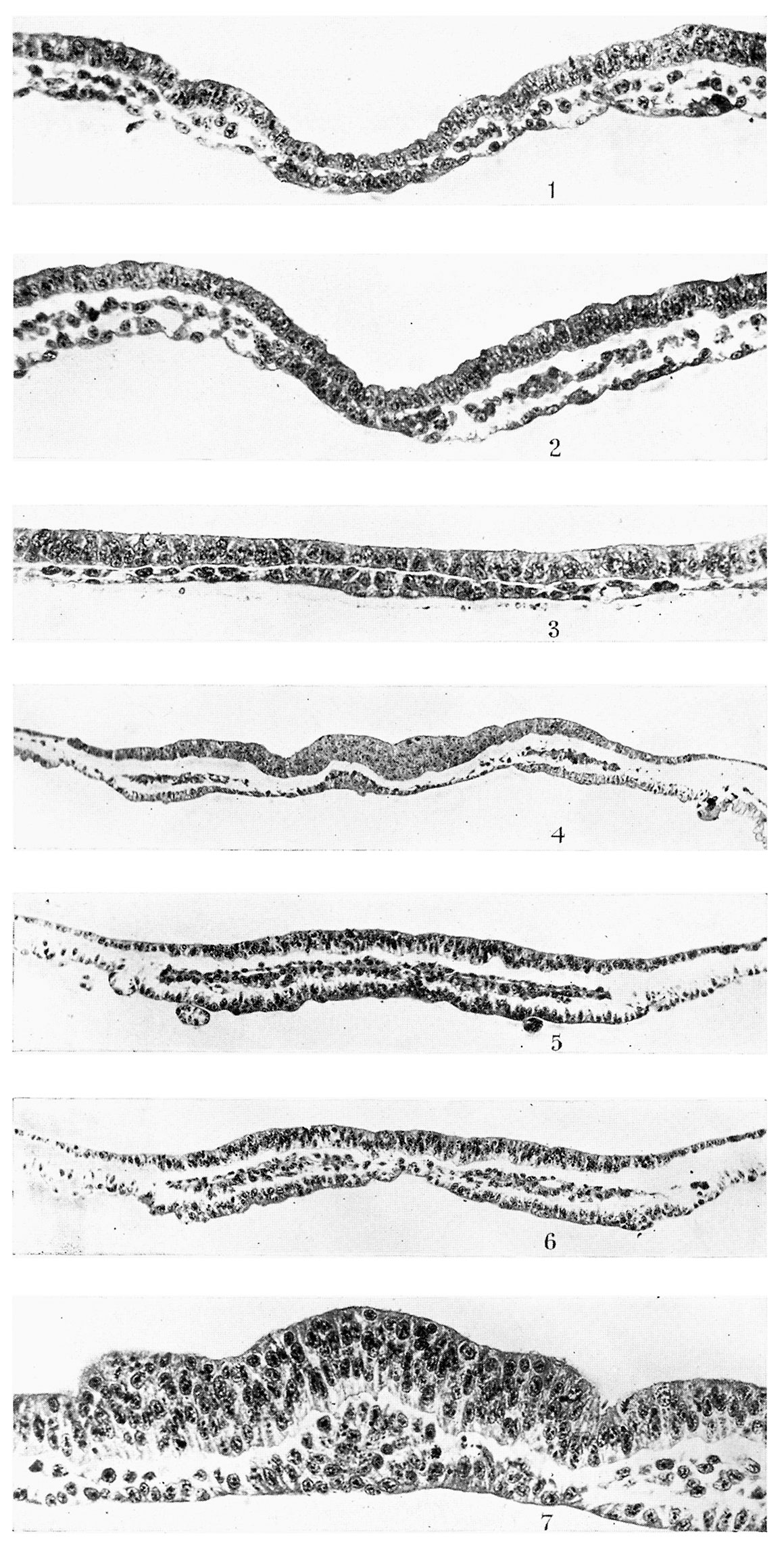
Plate 1
Fig. 1. T.S. (9.1.2.) of R 335, 8 days, primitive streak and head-process stage. x 330.
Fig. 2. T.S., 48th section behind fig. 1—same embryo as fig. 1. x 330.
Fig. 3. T.S. (4.2.2.) of R 352, 8 days 1 hour, ?I somite. x 330.
Fig. 4. T.S. (25.l.2.) of rabbit C’, 8 days, 3 somites. x 150.
Fig. 5. T.S., 22nd section of R 314, 8 days 17% hours, 4 somites. x 150.
Fig. 6. T.S., 28th section of same. x 150.
Fig. 7. T.S., 41st section of same. x 330.
Reference
Aasar YH. The history of the prochordal plate in the rabbit. (1931) J. Anat., 66(1):14-i3. PubMed 17104355
Cite this page: Hill, M.A. (2024, April 27) Embryology Aasar1931 plate1.jpg. Retrieved from https://embryology.med.unsw.edu.au/embryology/index.php/File:Aasar1931_plate1.jpg
- © Dr Mark Hill 2024, UNSW Embryology ISBN: 978 0 7334 2609 4 - UNSW CRICOS Provider Code No. 00098G
File history
Click on a date/time to view the file as it appeared at that time.
| Date/Time | Thumbnail | Dimensions | User | Comment | |
|---|---|---|---|---|---|
| current | 09:43, 9 October 2018 | | 1,160 × 2,287 (512 KB) | Z8600021 (talk | contribs) | |
| 09:41, 9 October 2018 |  | 1,614 × 2,530 (680 KB) | Z8600021 (talk | contribs) | ===Reference=== {{Ref-Aasar1931}} {{Footer}} Category:RabbitCategory:Historic EmbryologyCategory:1930's |
You cannot overwrite this file.
File usage
The following page uses this file: