File:Thyng1908 fig06.jpg
From Embryology

Size of this preview: 800 × 494 pixels. Other resolution: 879 × 543 pixels.
Original file (879 × 543 pixels, file size: 58 KB, MIME type: image/jpeg)
Fig. 6. Reconstruction from a human embryo of 13.6 mm
(H. E. C. 839). X 55 diams.
D. chol., ductus choledochus. D. cyst., ductus cysticus. D. hep., ductus hepaticus. D. panc. d., ductus pancreatis dorsalis. D. panc. v., ductus pancreatis ventralis. Panc. d., pancreas dorsale. Panc. d., pancreas ventrale. St., stomach.
Reference
Thyng FW. Models of the pancreas in embryos of the pig, rabbit, cat, and man. (1908) Amer. J Anat. 7(4): 489–503.
File history
Yi efo/eka'e gwa ebo wo le nyangagi wuncin ye kamina wunga tinya nan
| Gwalagizhi | Nyangagi | Dimensions | User | Comment | |
|---|---|---|---|---|---|
| current | 17:47, 23 Mayıs 2016 | | 879 × 543 (58 KB) | Z8600021 (talk | contribs) | |
| 17:47, 23 Mayıs 2016 | 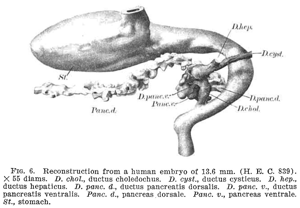 | 1,000 × 692 (99 KB) | Z8600021 (talk | contribs) | '''Fig. 6.''' Reconstruction from a human embryo of 13.6 mm. (H. E. C. 839). X 55 diams. D. choZ., ductus choledochus. D. cyszt, ductus cysticus. D. hep, ductus hepaticus. D. pane. d., ductus pancreatis dorsalis. D. pane. 12., ductus pancreatis ventra... |
You cannot overwrite this file.
File usage
The following page uses this file:


