File:Senior1919 fig03.jpg
From Embryology
Size of this preview: 800 × 505 pixels. Other resolution: 1,000 × 631 pixels.
Original file (1,000 × 631 pixels, file size: 92 KB, MIME type: image/jpeg)
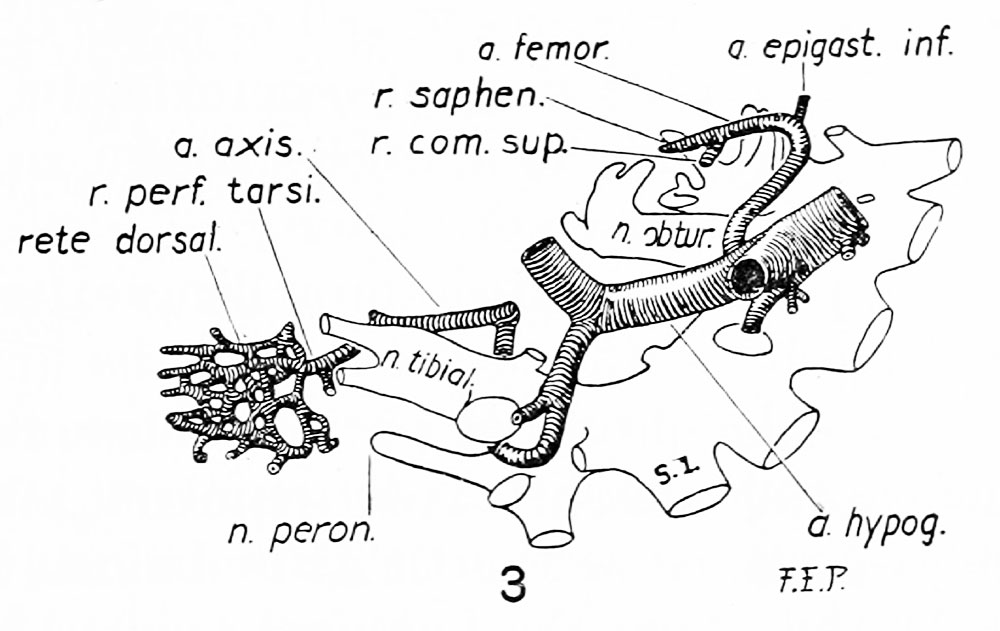
Fig. 3. Reconstruction showing the arteries of the right side of the pelvis, right thigh, leg, and dorsum of the foot in a human embryo of 12 mm
(M.E. C.,H. 14). Medial aspect. X 20 diams,
Reference
Senior HD. The development of the arteries of the human lower extremity. (1919) Amer. J Anat. 22:1-11.
Cite this page: Hill, M.A. (2026, April 18) Embryology Senior1919 fig03.jpg. Retrieved from https://embryology.med.unsw.edu.au/embryology/index.php/File:Senior1919_fig03.jpg
- © Dr Mark Hill 2026, UNSW Embryology ISBN: 978 0 7334 2609 4 - UNSW CRICOS Provider Code No. 00098G
File history
Yi efo/eka'e gwa ebo wo le nyangagi wuncin ye kamina wunga tinya nan
| Gwalagizhi | Nyangagi | Dimensions | User | Comment | |
|---|---|---|---|---|---|
| current | 09:25, 27 October 2016 | | 1,000 × 631 (92 KB) | Z8600021 (talk | contribs) | |
| 09:25, 27 October 2016 |  | 2,500 × 2,099 (500 KB) | Z8600021 (talk | contribs) |
You cannot overwrite this file.
File usage
The following 2 pages use this file: