File:Sea urchin SEM01.jpg
Original file (1,000 × 712 pixels, file size: 95 KB, MIME type: image/jpeg)
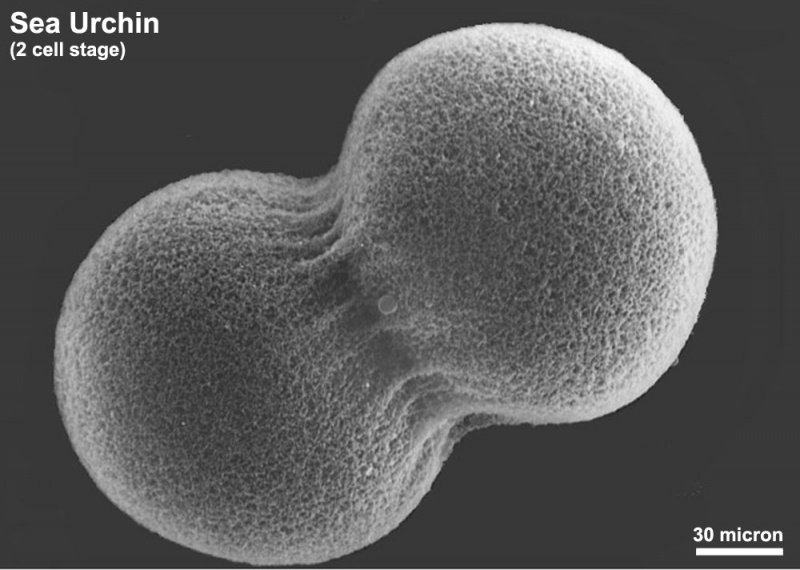
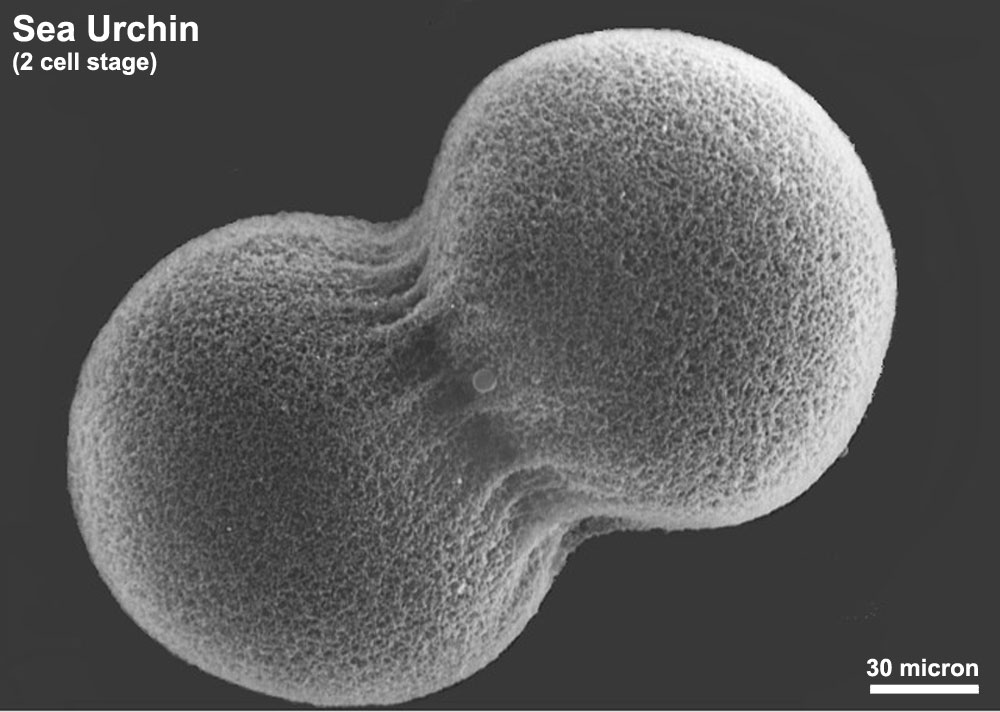
Sea Urchin (2 cell stage)
Scanning electron microscope image of Lytechinus pictus [sea urchin] embryo at the 2-cell stage. Fertilization envelope has been removed to reveal the cells covered with a dense meshwork of the hyaline layer, in which microvilli are embedded.
Class Echinoidea - Superorder Echinacea - OrderTemnopleuroida - Lytechinus pictus
- Links: Sea Urchin Development
Image Source: Contributed by Dartmouth College Electron Microscope Facility special thanks to Chuck Daghlian and Louisa Howard. Gallery. Original images may have been altered in size contrast and labelling. (These images are in the public domain)
JEOL 35C SEM Evelyn Spiegel, Louisa Howard
File history
Yi efo/eka'e gwa ebo wo le nyangagi wuncin ye kamina wunga tinya nan
| Gwalagizhi | Nyangagi | Dimensions | User | Comment | |
|---|---|---|---|---|---|
| current | 15:22, 1 Haziran 2011 | | 1,000 × 712 (95 KB) | S8600021 (talk | contribs) | ==Sea Urchin (2 cell stage)== Scanning electron microscope image of Lytechinus pictus [sea urchin] embryo at the 2-cell stage. Fertilization envelope has been removed to reveal the cells covered with a dense meshwork of the hyaline layer, in which microv |
You cannot overwrite this file.
File usage
The following 2 pages use this file: