File:Reprogramming MEF into ES-like cells 01.jpg
Original file (600 × 888 pixels, file size: 145 KB, MIME type: image/jpeg)
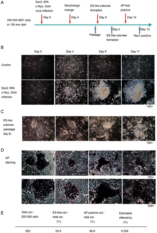
Reprogramming of genetically unmodified MEF into ES-like cells
(A) Outline of the MEF reprogramming protocol. 2.5×10⁵ MEF cells were plated in two 100 mm dishes and were infected with Sox2, Klf4, c-Myc and Oct3/4 virus. Cells in dish 1(blue arrow) were passage on day 8 after infection and ES-like colonies form 4 days later. Cells in dish 2 (red arrow) were AP staining positive on day 14 and the colonies were counted. (B) Significant morphology changes observed on day 4 and ES-like colonies form on day 9 after MEF cells were infected with Sox2, Klf4, c-Myc and Oct3/4 virus. (C) Morphology of ES-like colonies on day 6 after passage. (D) AP positive colonies on day 14 post infection. (E) Estimated reprogramming efficiency of genetically unmodified MEF into AP positive ES-like cells 16 days post infection.
File:Reprogramming MEF into ES-like cells 02.jpg
(F) ES-like cell lines were established using the nonselective approach. Representative colonies from passage 6 cells were stained with anti-Rex1, Sox2 and SSEA1 antibodies and images were acquired through a Leica confocal system.
Original file name: Cr200792f1a.jpg
Reference
<pubmed>17971807</pubmed>| Cell Research
Reprinted by permission from Macmillan Publishers Ltd
Licensee: Mark A Hill License Date: May 24, 2011 License Number: 2675351286106 Publication: Cell Research Title: Direct generation of ES-like cells from unmodified mouse embryonic fibroblasts by Oct4/Sox2/Myc/Klf4 Type Of Use: post on the internet
http://s100.copyright.com/CustomerAdmin/PLF.jsp?lID=2011051_1306242206106
File history
Click on a date/time to view the file as it appeared at that time.
| Date/Time | Thumbnail | Dimensions | User | Comment | |
|---|---|---|---|---|---|
| current | 23:08, 24 May 2011 | | 600 × 888 (145 KB) | S8600021 (talk | contribs) | ==Reprogramming of genetically unmodified MEF into ES-like cells== (A) Outline of the MEF reprogramming protocol. 2.5×10⁵ MEF cells were plated in two 100 mm dishes and were infected with Sox2, Klf4, c-Myc and Oct3/4 virus. Cells in dish 1(blue arrow) w |
You cannot overwrite this file.
File usage
The following page uses this file: