File:Odgers1941 text-fig01.jpg

Original file (1,000 × 995 pixels, file size: 51 KB, MIME type: image/jpeg)
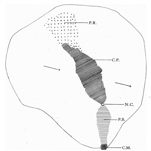
Text-Fig.1. Dorsal aspect of the embryonal shield
A projection of the dorsal aspect of the embryonal shield on the flat (x 75), drawn to scale. C.M. cloacal membrane. P.S. primitive streak. N.C. neurenteric canal. C.P. chorda plate. P.R.(with coarse dots) prochordal plate.Thet at the cranial end of the chord plate indicates the site of the remains of the chorda canal. The arrows indicate the plane of section.
Reference
Odgers PN. A presomite human embryo with a neurenteric canal (embryo R.S.). (1941) J. Anat., 75(4): 381-388.3. PMID 17104868
Cite this page: Hill, M.A. (2024, June 5) Embryology Odgers1941 text-fig01.jpg. Retrieved from https://embryology.med.unsw.edu.au/embryology/index.php/File:Odgers1941_text-fig01.jpg
- © Dr Mark Hill 2024, UNSW Embryology ISBN: 978 0 7334 2609 4 - UNSW CRICOS Provider Code No. 00098G
File history
Click on a date/time to view the file as it appeared at that time.
| Date/Time | Thumbnail | Dimensions | User | Comment | |
|---|---|---|---|---|---|
| current | 16:53, 22 October 2017 | | 1,000 × 995 (51 KB) | Z8600021 (talk | contribs) | |
| 16:52, 22 October 2017 | 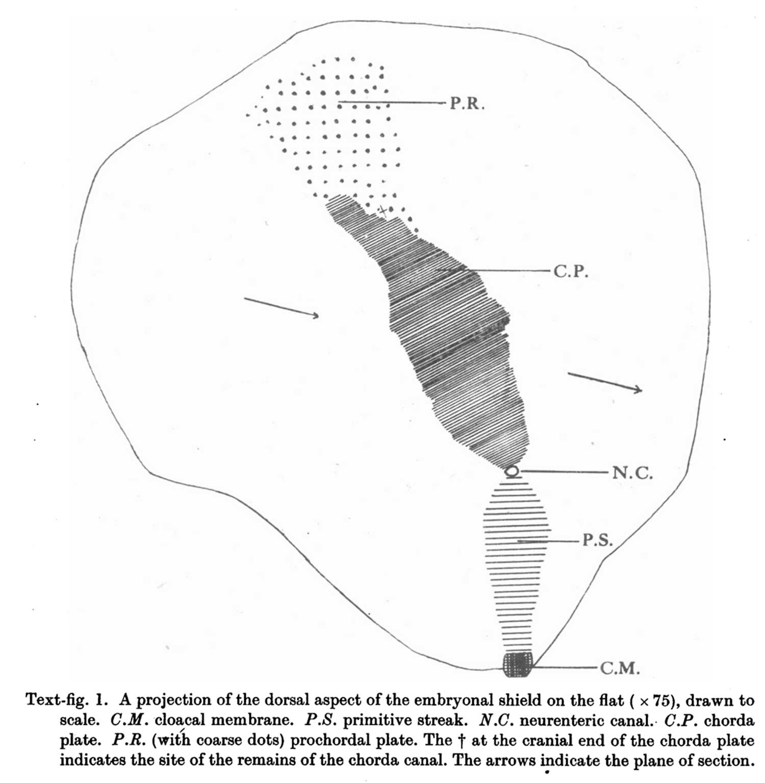 | 1,500 × 1,534 (104 KB) | Z8600021 (talk | contribs) | ===Reference=== {{Ref-Odgers1941}} {{Footer}} |
You cannot overwrite this file.
File usage
The following page uses this file: