File:Odgers1939-fig03.jpg
From Embryology
Size of this preview: 369 × 600 pixels. Other resolution: 550 × 894 pixels.
Original file (550 × 894 pixels, file size: 154 KB, MIME type: image/jpeg)
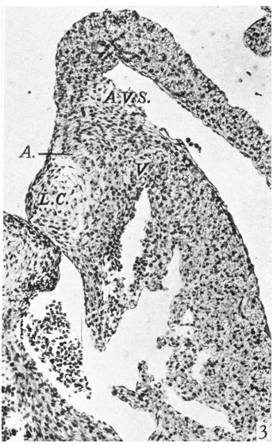
Fig. 3. A section through the left lateral cusp in a 17-5 mm. embryo
( x 109) to show in contrast the absence of any angulation of the sulcus, A.V.S., while the left lateral cushion, L.0., maintains its original shape and relations to A., auricular muscle and to V., ventricular muscle.
File history
Yi efo/eka'e gwa ebo wo le nyangagi wuncin ye kamina wunga tinya nan
| Gwalagizhi | Nyangagi | Dimensions | User | Comment | |
|---|---|---|---|---|---|
| current | 15:00, 15 November 2015 | | 550 × 894 (154 KB) | Z8600021 (talk | contribs) |
You cannot overwrite this file.
File usage
The following 3 pages use this file: