File:Mouse-embryo granzyme G.jpg

Original file (899 × 1,000 pixels, file size: 90 KB, MIME type: image/jpeg)
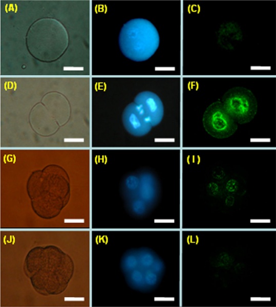
Early Embryo Granzyme G Expression (mouse)
Whole-mount embryo in situ hybridization (ISH) using a FITC-conjugated granzyme G oligonucleotide probe at different stages of normal mouse development.
- (A to C) One-cell stage mouse embryos.
- (D to F) Two-cell stage mouse embryos.
- (G to I) Four-cell stage mouse embryos.
- (J to K) Eight-cell stage mouse embryos.
- The left panels (A, D, G, and J) show embryos photographed under phase contrast imaging.
- The middle panels (B, E, H, and K) show embryos stained with Hoechst 33342 for DNA localization (blue) under fluorescence microscope observation.
- The right panels (C, F, I, and L) show embryos hybridized with a granzyme G oligonucleotide probe (green) under confocal microscopic observation.
Scale bar: 30 μm.
- "The expression of members of the granzyme gene family of proteins (granzymes A-H, K, M), which encode serine proteases, has been documented in the secretory granules of cytolytic T lymphocyte lines [12]. Granzymes D, E, F, and G have also been shown to be expressed at gestation in the mouse uterus during the process of decidualization, in which rapid uterine cell growth and differentiation occurs [13]."
Reference
<pubmed>20704734</pubmed>| BMC Dev Biol.
Tsai et al. BMC Developmental Biology 2010 10:88 doi:10.1186/1471-213X-10-88
© 2010 Tsai et al; licensee BioMed Central Ltd. This is an Open Access article distributed under the terms of the Creative Commons Attribution License (http://creativecommons.org/licenses/by/2.0), which permits unrestricted use, distribution, and reproduction in any medium, provided the original work is properly cited.
File history
Click on a date/time to view the file as it appeared at that time.
| Date/Time | Thumbnail | Dimensions | User | Comment | |
|---|---|---|---|---|---|
| current | 13:54, 19 October 2010 | | 899 × 1,000 (90 KB) | S8600021 (talk | contribs) | ==Early Embryo Granzyme G Expression (mouse)== Whole-mount embryo in situ hybridization (ISH) using a FITC-conjugated granzyme G oligonucleotide probe at different stages of normal mouse development. (A to C) One-cell stage mouse embryos. (D to F) Tw |
You cannot overwrite this file.
File usage
There are no pages that use this file.