File:Morgan 1925 fig32.jpg
From Embryology
Size of this preview: 501 × 599 pixels. Other resolution: 836 × 1,000 pixels.
Original file (836 × 1,000 pixels, file size: 194 KB, MIME type: image/jpeg)
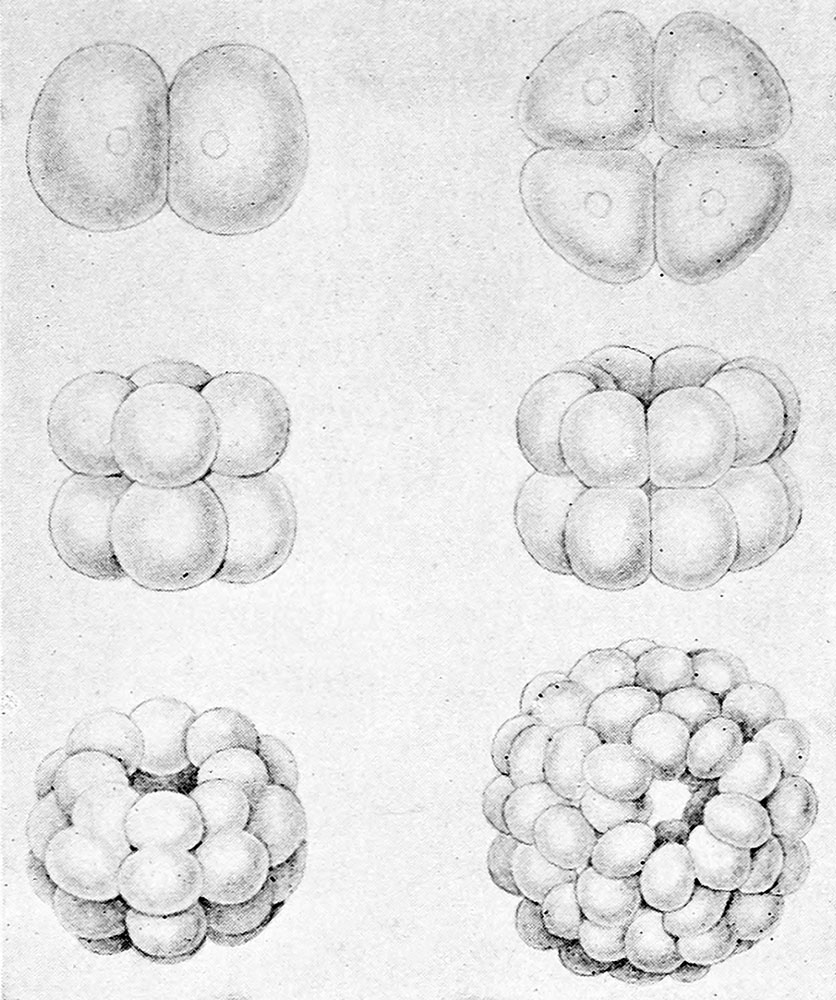
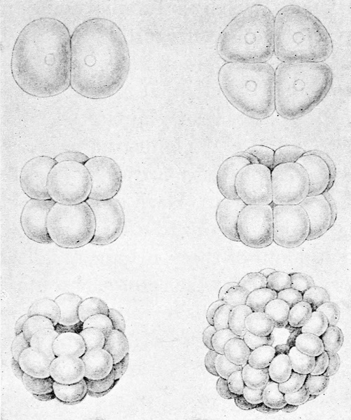
Fig. 32. Diagram showing the segmentation of an egg into two, four, eight cells
The cells become arranged over the surface of a sphere whose interior is filled with fluid. (After Selenka.)
| Historic Disclaimer - information about historic embryology pages |
|---|
 |
Reference
Morgan, T. H. (1925). Evolution and genetics. Princeton: Princeton University Press.
Cite this page: Hill, M.A. (2024, June 3) Embryology Morgan 1925 fig32.jpg. Retrieved from https://embryology.med.unsw.edu.au/embryology/index.php/File:Morgan_1925_fig32.jpg
- © Dr Mark Hill 2024, UNSW Embryology ISBN: 978 0 7334 2609 4 - UNSW CRICOS Provider Code No. 00098G
File history
Click on a date/time to view the file as it appeared at that time.
| Date/Time | Thumbnail | Dimensions | User | Comment | |
|---|---|---|---|---|---|
| current | 09:08, 23 October 2014 | | 836 × 1,000 (194 KB) | Z8600021 (talk | contribs) | |
| 09:06, 23 October 2014 | 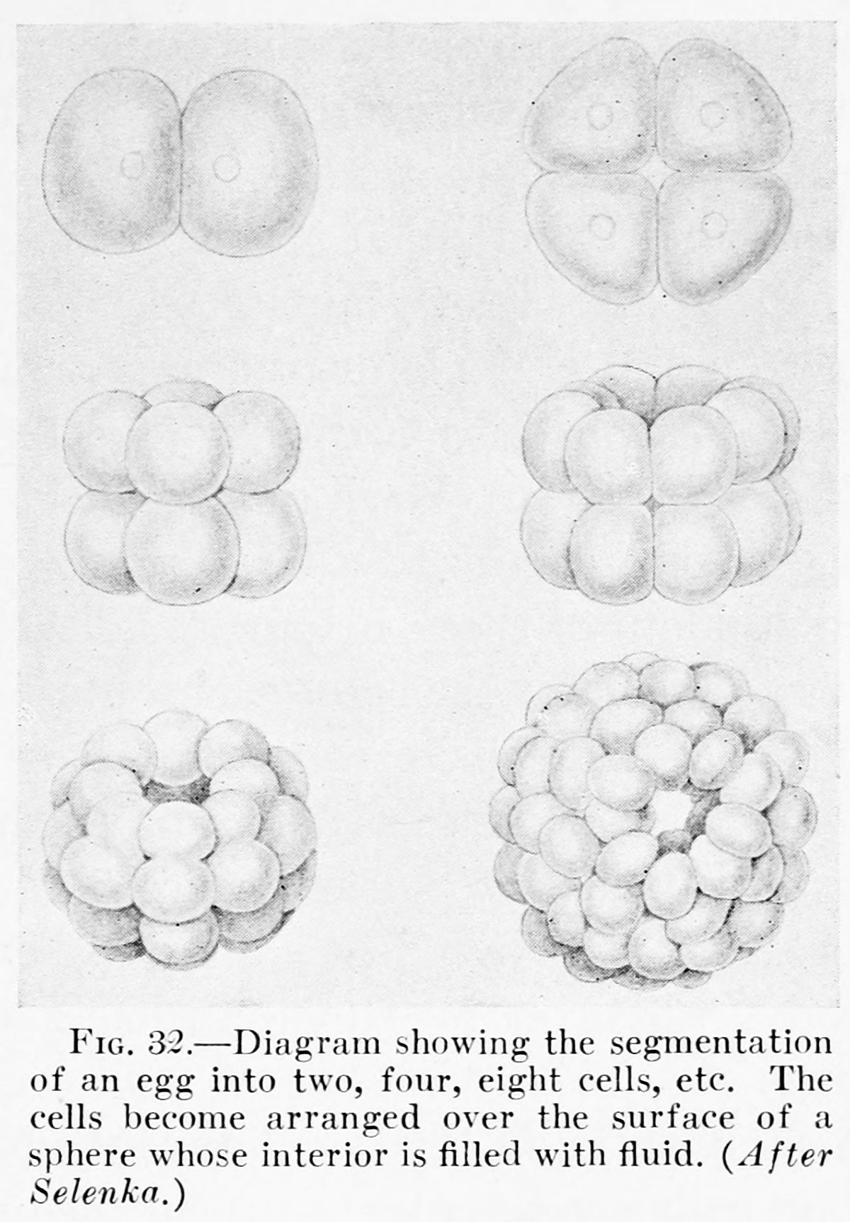 | 1,200 × 1,725 (303 KB) | Z8600021 (talk | contribs) |
You cannot overwrite this file.
File usage
The following 2 pages use this file: