File:Lewis1903 plate01L.jpg
Original file (1,280 × 1,822 pixels, file size: 246 KB, MIME type: image/jpeg)
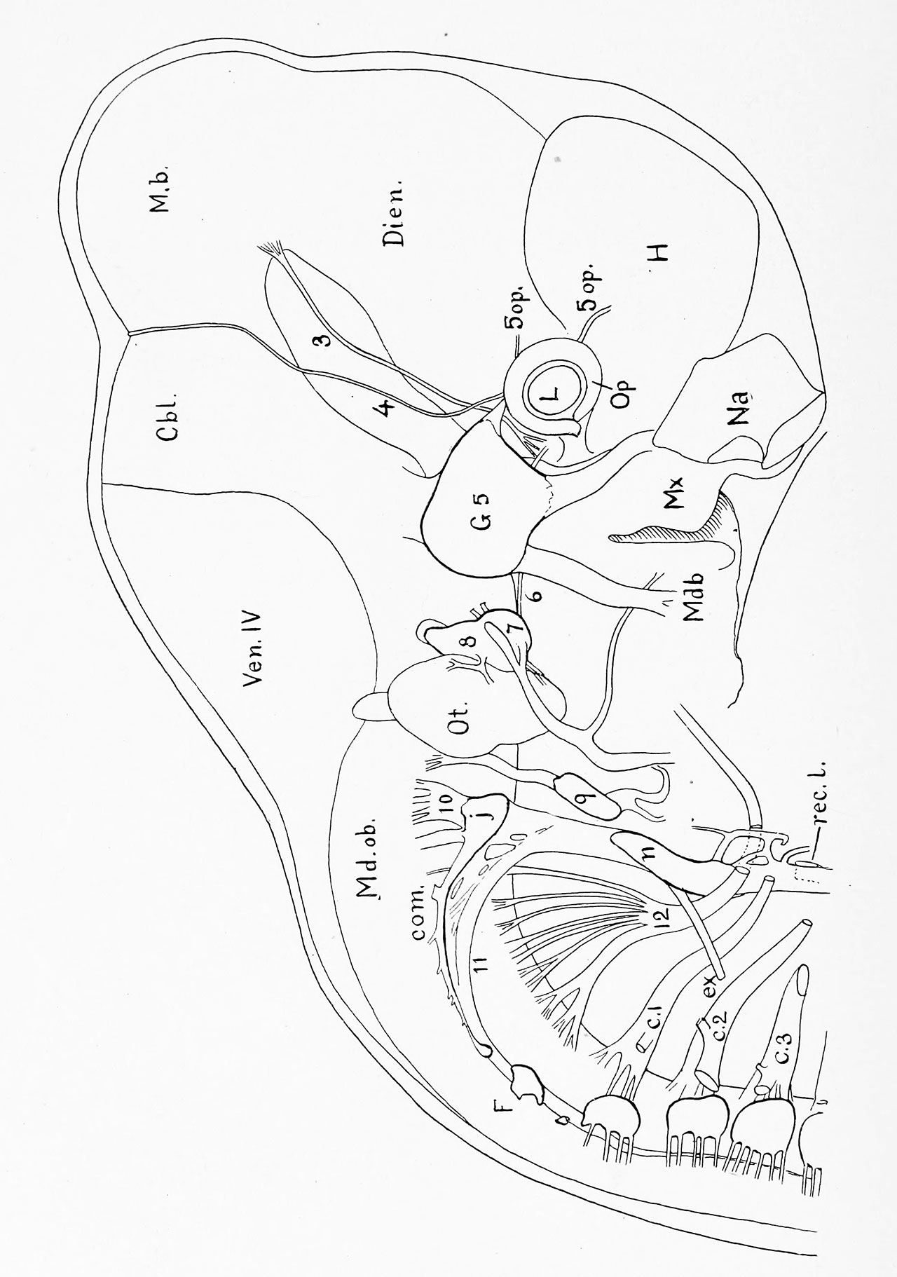
Plate I Pig Embryo of 12.0 mm
Reconstruction from transverse sections, Series 5.
To show especially the cephalic nerves, c. 1, c. 2, c. 3, Cervical nerves. Cbl, Cerebellum, com. Ganglionic commissure. Dien, Diencephalon. e.i'. External branch of the spinal accessory nerve. F, Froriep's ganglion. Gf. 5, Gasserian ganglion. H, Cerebral hemisphere. ;, Jugular ganglion. L, Lens. M. b, Mid-brain. Mdb., Mandibular process. Md. ob, Medulla oblongata. Mic, Maxillary process, n, Ganglion nodosum. Na, Nasal pit. Op, Optic cup. Ot, Otocyst. Rec. I, Recurrent laryngeal nerve. Yen. IV, Roof of fourth ventricle. 3, Oculomotor nerve. 4, Trochlear nerve. Sop, Branches of the ophthalmic division of the trigeminal nerve. 6, Abducens nerve. 7, Geniciilate ganglion of the facial nerve. 8, Vestibular ganglion. 9, Petrosal ganglion. 10, Vagus nerve. 11, Spinal accessory nerve. 12, Hypoglossal nerve.
x 20 diams.
Reference
Lewis FT. The gross anatomy of a 12 mm pig. (1903) Amer. J Anat. 2: 221-225.
Cite this page: Hill, M.A. (2026, September 15) Embryology Lewis1903 plate01L.jpg. Retrieved from https://embryology.med.unsw.edu.au/embryology/index.php/File:Lewis1903_plate01L.jpg
- © Dr Mark Hill 2026, UNSW Embryology ISBN: 978 0 7334 2609 4 - UNSW CRICOS Provider Code No. 00098G
File history
Yi efo/eka'e gwa ebo wo le nyangagi wuncin ye kamina wunga tinya nan
| Gwalagizhi | Nyangagi | Dimensions | User | Comment | |
|---|---|---|---|---|---|
| current | 13:24, 2 August 2019 | | 1,280 × 1,822 (246 KB) | Z8600021 (talk | contribs) | |
| 13:24, 2 August 2019 |  | 1,860 × 2,647 (461 KB) | Z8600021 (talk | contribs) | ||
| 13:23, 2 August 2019 |  | 2,454 × 3,641 (531 KB) | Z8600021 (talk | contribs) | labels for plate 1 |
You cannot overwrite this file.
File usage
The following page uses this file:


