File:Kindred1933 fig05.jpg
From Embryology

Size of this preview: 643 × 599 pixels. Other resolution: 1,000 × 932 pixels.
Original file (1,000 × 932 pixels, file size: 278 KB, MIME type: image/jpeg)
Fig.5 Drawing of model of embryo
The amnion is cut at the region of junction with the embryonic area. Dorsal view. The model is 100 times the size of the embryo. The numbers on the line below the model refer to the planes of the sections shown in plate 1.
Reference
Kindred JE. A human embryo of the pre-somite period from the uterine tube. (1933) Amer. J Anat. 53: 221-241.
Cite this page: Hill, M.A. (2026, October 2) Embryology Kindred1933 fig05.jpg. Retrieved from https://embryology.med.unsw.edu.au/embryology/index.php/File:Kindred1933_fig05.jpg
- © Dr Mark Hill 2026, UNSW Embryology ISBN: 978 0 7334 2609 4 - UNSW CRICOS Provider Code No. 00098G
File history
Yi efo/eka'e gwa ebo wo le nyangagi wuncin ye kamina wunga tinya nan
| Gwalagizhi | Nyangagi | Dimensions | User | Comment | |
|---|---|---|---|---|---|
| current | 17:59, 27 November 2016 | | 1,000 × 932 (278 KB) | Z8600021 (talk | contribs) | |
| 17:59, 27 November 2016 | 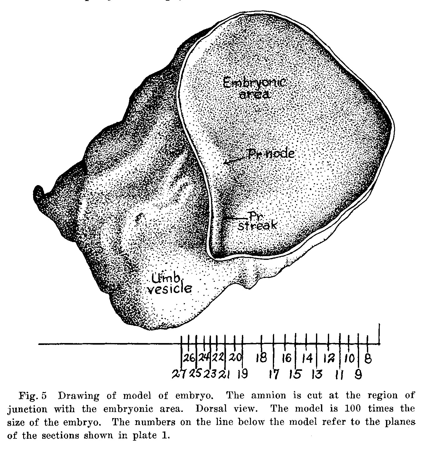 | 1,363 × 1,439 (557 KB) | Z8600021 (talk | contribs) |
You cannot overwrite this file.
File usage
The following page uses this file:


