File:Hypospadia 3D ultrasound 01.jpg

Original file (1,150 × 497 pixels, file size: 87 KB, MIME type: image/jpeg)
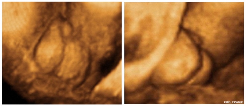
Penoscrotal Hypospadia 3D Ultrasound
Ultrasonography in rendering mode, at GA 33 weeks, with short penis and with evidence of testicles inside a bifid scrotum.
Reference
<pubmed>23304621</pubmed>| PMC3530751 | Case Rep Urol.
Copyright
© 2012 Lívia Teresa Moreira Rios et al. This is an open access article distributed under the Creative Commons Attribution License, which permits unrestricted use, distribution, and reproduction in any medium, provided the original work is properly cited. Figure 2 http://www.hindawi.com/journals/criu/2012/142814/fig2/
Cite this page: Hill, M.A. (2026, October 4) Embryology Hypospadia 3D ultrasound 01.jpg. Retrieved from https://embryology.med.unsw.edu.au/embryology/index.php/File:Hypospadia_3D_ultrasound_01.jpg
- © Dr Mark Hill 2026, UNSW Embryology ISBN: 978 0 7334 2609 4 - UNSW CRICOS Provider Code No. 00098G
File history
Yi efo/eka'e gwa ebo wo le nyangagi wuncin ye kamina wunga tinya nan
| Gwalagizhi | Nyangagi | Dimensions | User | Comment | |
|---|---|---|---|---|---|
| current | 08:54, 17 Mayıs 2015 | | 1,150 × 497 (87 KB) | Z8600021 (talk | contribs) | ==Penoscrotal Hypospadia 3D Ultrasound== Ultrasonography in rendering mode, at ((GA)) 33 weeks, with short penis and with evidence of testicles inside a bifid scrotum. Figure 2 http://www.hindawi.com/journals/criu/2012/142814/fig2/ |
You cannot overwrite this file.
File usage
The following 2 pages use this file: