File:Human placental villi cartoon 01.jpg
From Embryology

Size of this preview: 800 × 489 pixels. Other resolution: 1,084 × 663 pixels.
Original file (1,084 × 663 pixels, file size: 142 KB, MIME type: image/jpeg)
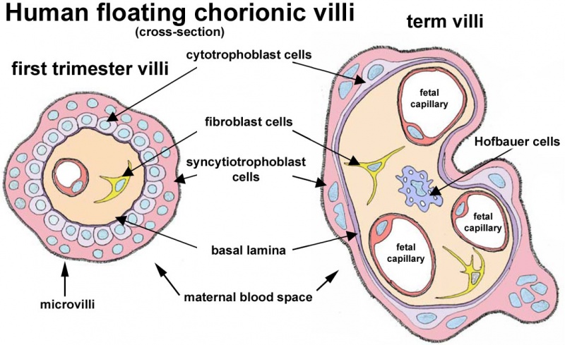
Human Floating Chorionic Villi
A figure showing the changes in placental villi between early (first trimester) and late (third trimester) placental development.
Villi Structure:
- syncytiotrophoblast and cytotrophoblast change their cellular organisation.
- Capillaries increase within the villi, at term nearly the entire villi is occupied by capillaries with only minimal surrounding connective tissue.
- Hofbauer cells are abundant in villi in both first and second trimesters (though only shown in the term villi). Later Hofbauer cells may have a different embryonic origin.
- Links: placental villi | syncytiotrophoblast | cytotrophoblast | Hofbauer cells
Reference
Figure based Fig 1.B Malassiné et al. Retrovirology 2008 5:6 doi:10.1186/1742-4690-5-6.
Cite this page: Hill, M.A. (2026, October 11) Embryology Human placental villi cartoon 01.jpg. Retrieved from https://embryology.med.unsw.edu.au/embryology/index.php/File:Human_placental_villi_cartoon_01.jpg
- © Dr Mark Hill 2026, UNSW Embryology ISBN: 978 0 7334 2609 4 - UNSW CRICOS Provider Code No. 00098G
File history
Yi efo/eka'e gwa ebo wo le nyangagi wuncin ye kamina wunga tinya nan
| Gwalagizhi | Nyangagi | Dimensions | User | Comment | |
|---|---|---|---|---|---|
| current | 13:13, 3 Haziran 2012 | | 1,084 × 663 (142 KB) | Z8600021 (talk | contribs) | ==Human Floating Chorionic Villi== ===Reference=== Figure based Fig 1. Malassiné et al. [http://www.retrovirology.com/content/5/1/6 Retrovirology] 2008 5:6 doi:10.1186/1742-4690-5-6. |
You cannot overwrite this file.