File:Hertig1946b fig26a.jpg
Original file (800 × 646 pixels, file size: 131 KB, MIME type: image/jpeg)
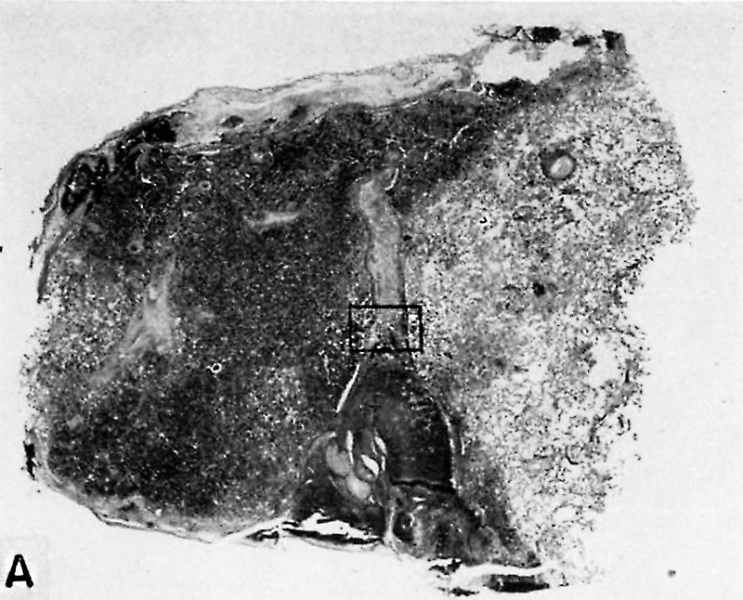
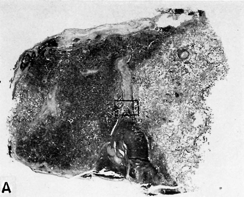
Fig. 26. A human placenta at term
To show the cystic degeneration occurring in the “fibrinoid" of a septum separating two adjacent cotyledons. B. L.-i. H., S-34-581.
A. A low power view of two cotyledons separated by a cytotrophoblastic septum whose "fibrinoid” has undergone cystic degeneration. The resultant hematoma at the base has disrupted the blood supply of the cotyledon at the left and caused infarction but has spared the one to the right. X5.
References
Hertig AT. lnvolution of tissues in fetal life: a review. (1946) Anat. Rec. 94: 96-116.
Cite this page: Hill, M.A. (2026, September 28) Embryology Hertig1946b fig26a.jpg. Retrieved from https://embryology.med.unsw.edu.au/embryology/index.php/File:Hertig1946b_fig26a.jpg
- © Dr Mark Hill 2026, UNSW Embryology ISBN: 978 0 7334 2609 4 - UNSW CRICOS Provider Code No. 00098G
File history
Yi efo/eka'e gwa ebo wo le nyangagi wuncin ye kamina wunga tinya nan
| Gwalagizhi | Nyangagi | Dimensions | User | Comment | |
|---|---|---|---|---|---|
| current | 10:46, 8 August 2017 | | 800 × 646 (131 KB) | Z8600021 (talk | contribs) |
You cannot overwrite this file.
File usage
The following 2 pages use this file: