File:Hertig1946b fig17.jpg

Original file (800 × 1,155 pixels, file size: 161 KB, MIME type: image/jpeg)
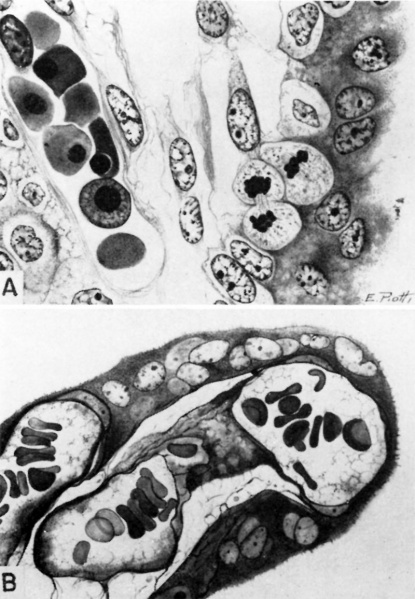
Fig. 17. Human Chorionic Villus
A. A cytologic drawing of a section from a 30-day human chorionic villus (Fig. 4 from Wislocki and Bennett, American Journal of Anatomy). The thick syncytium is seen covering the actively growing Langhans epithelium. The loose immature connective tissue contains a thin-walled capillary within which are seen fetal blood cells. Note that the erythroblasts are frequently nucleated. Iron hematoxylin, X1600.
B. A histologic drawing of a mature human chorionic villus (Fig. 17 from Wislocki and Bennett, American Journal of Anatomy). The syncytium is now much thinner, especially overlying the dilated fetal capillaries. Langhans cells are absent in this section although mature villi still contain a few scattered ones. The stroma is dense, the capillaries numerous, the latter containing many non nucleated fetal erythroblasts. Mallory’s connective tissue stain, X1600.
References
Hertig AT. lnvolution of tissues in fetal life: a review. (1946) Anat. Rec. 94: 96-116.
Cite this page: Hill, M.A. (2026, September 25) Embryology Hertig1946b fig17.jpg. Retrieved from https://embryology.med.unsw.edu.au/embryology/index.php/File:Hertig1946b_fig17.jpg
- © Dr Mark Hill 2026, UNSW Embryology ISBN: 978 0 7334 2609 4 - UNSW CRICOS Provider Code No. 00098G
File history
Yi efo/eka'e gwa ebo wo le nyangagi wuncin ye kamina wunga tinya nan
| Gwalagizhi | Nyangagi | Dimensions | User | Comment | |
|---|---|---|---|---|---|
| current | 08:58, 8 August 2017 | | 800 × 1,155 (161 KB) | Z8600021 (talk | contribs) |
You cannot overwrite this file.
File usage
The following page uses this file: