File:Hertig1946b fig04a.jpg

Original file (800 × 667 pixels, file size: 128 KB, MIME type: image/jpeg)
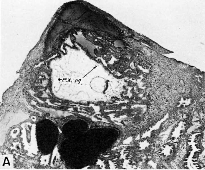
Fig. 4A. A human ovum of approximately 13.5 days of age
The hemorrhagic closing cap is seen above the ovum. The chorion possesses simple unbranched villi. The chorionic cavity contains an eccentrically situated embryo (see fig. 4B for details of the latter) and the remnants of the exocoelomic membrane (EX. M). The endometrium shows an early decidual reaction about the'ovum. The gland below the ovum contains extravasated blood. Note large sinusoid in the endometrium to the left of the ovum. Carnegie 7801, section 12-1-1, X35.
References
Hertig AT. lnvolution of tissues in fetal life: a review. (1946) Anat. Rec. 94: 96-116.
Cite this page: Hill, M.A. (2026, April 18) Embryology Hertig1946b fig04a.jpg. Retrieved from https://embryology.med.unsw.edu.au/embryology/index.php/File:Hertig1946b_fig04a.jpg
- © Dr Mark Hill 2026, UNSW Embryology ISBN: 978 0 7334 2609 4 - UNSW CRICOS Provider Code No. 00098G
File history
Yi efo/eka'e gwa ebo wo le nyangagi wuncin ye kamina wunga tinya nan
| Gwalagizhi | Nyangagi | Dimensions | User | Comment | |
|---|---|---|---|---|---|
| current | 16:47, 7 August 2017 | | 800 × 667 (128 KB) | Z8600021 (talk | contribs) |
You cannot overwrite this file.
File usage
The following page uses this file: