File:Hertig1945d fig05.jpg
Original file (1,280 × 412 pixels, file size: 84 KB, MIME type: image/jpeg)
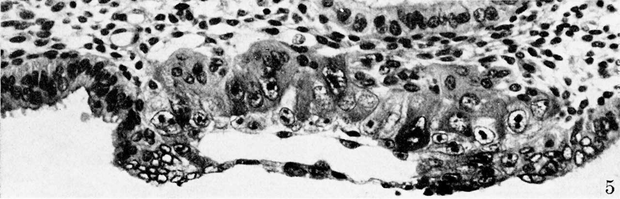
Fig. 5. A section of a 7.5-day ovum
(same specimen as figs. 2-4) showing a more advanced stage of mesoblastic formation. Note the two, nearly detached mesoblastic cells delaminating from, but still lightly attached to the trophoblast at the embryonic pole of the ovum. Carnegie 8020, section 6-4-3, X 300 (plus).
Fig 1 Carnegie No. 8225
Fig 2 Carnegie No. 8020
Fig 3 Carnegie No. 8020
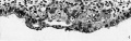
Fig 4 Carnegie No. 8020

Fig 5 Carnegie No. 8020
Reference
Hertig AT. On the development of the amnion and exocoelomic membrane in the previllous human ovum. (1945) Yale J Biol Med. 18:107-15. PubMed 21007544
Cite this page: Hill, M.A. (2024, May 23) Embryology Hertig1945d fig05.jpg. Retrieved from https://embryology.med.unsw.edu.au/embryology/index.php/File:Hertig1945d_fig05.jpg
- © Dr Mark Hill 2024, UNSW Embryology ISBN: 978 0 7334 2609 4 - UNSW CRICOS Provider Code No. 00098G
File history
Click on a date/time to view the file as it appeared at that time.
| Date/Time | Thumbnail | Dimensions | User | Comment | |
|---|---|---|---|---|---|
| current | 15:18, 24 October 2017 | 1,280 × 412 (84 KB) | Z8600021 (talk | contribs) |
You cannot overwrite this file.
File usage
The following 7 pages use this file: