File:Hertig1945d fig01.jpg

Original file (1,280 × 395 pixels, file size: 96 KB, MIME type: image/jpeg)
Fig. 1. The middle cross section of an ovum not over 7.5 days of age
Above the somewhat eccentrically placed germ-disk is a barely discernible cleft—the amniotic cavity. Above the latter are a few dark, flattened amniogenic cells de-lamiznating in situ from the adjacent trophoblast. Toward the left of the chorionic cavity, a few primitive mesoblastic cells are seen which likewise arise from trophoblast by in situ delamination. Carnegie No. 8225, section 20-5-5, X 300 (minus).
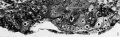
Fig 1 Carnegie No. 8225
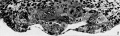
Fig 2 Carnegie No. 8020
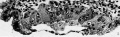
Fig 3 Carnegie No. 8020
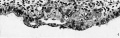
Fig 4 Carnegie No. 8020
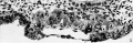
Fig 5 Carnegie No. 8020
Reference
Hertig AT. On the development of the amnion and exocoelomic membrane in the previllous human ovum. (1945) Yale J Biol Med. 18:107-15. PubMed 21007544
Cite this page: Hill, M.A. (2024, May 23) Embryology Hertig1945d fig01.jpg. Retrieved from https://embryology.med.unsw.edu.au/embryology/index.php/File:Hertig1945d_fig01.jpg
- © Dr Mark Hill 2024, UNSW Embryology ISBN: 978 0 7334 2609 4 - UNSW CRICOS Provider Code No. 00098G
File history
Click on a date/time to view the file as it appeared at that time.
| Date/Time | Thumbnail | Dimensions | User | Comment | |
|---|---|---|---|---|---|
| current | 15:17, 24 October 2017 | 1,280 × 395 (96 KB) | Z8600021 (talk | contribs) |
You cannot overwrite this file.
File usage
The following 7 pages use this file: