File:HansonAnson1962 fig03.jpg
Original file (1,280 × 612 pixels, file size: 215 KB, MIME type: image/jpeg)
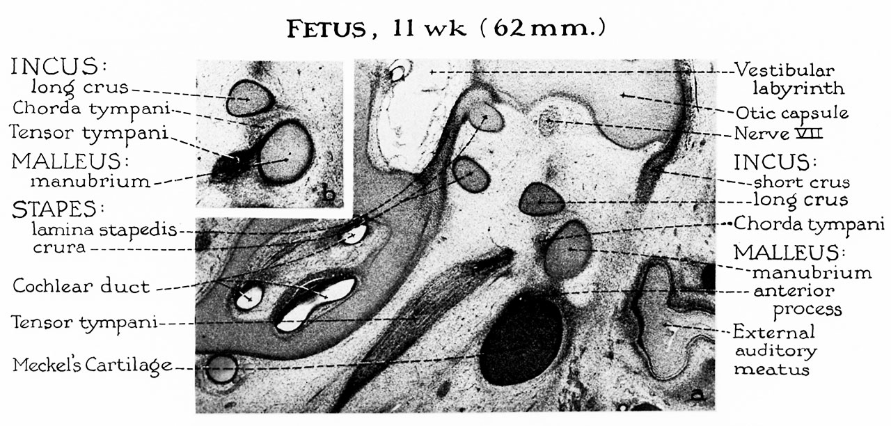
Fig. 3. Fetus 11 week 62 mm
a, In less than a 3-week period, the ossicles have doubled in size. However, they remain embedded in the loose mesenchyme of the tympanic cavity. Meckel’s cartilage continues to grow at a rate comparable to that of the malleus; as a result, in the fetus of 11 weeks the anterior process is dwarfed by this relatively large cartilaginous bar. The lamina stapedis is distinguishable from the remainder of the otic capsule. The otic (endolymphatic) labyrinth is formed, but the future periotic (perilymphatic) labyrinth is represented by reticular tissue (except when an early space forecasts the vestibule).
b, The newly formed tensor tympani muscle inserts into the manubrium of the malleus below the level of the chorda tympani, and below that of the lateral process (not shown). This projection is a cartilaginous outgrowth from the upper portion of the lateral aspect of the manubrium.,
Reference
Hanson JR. and Anson BJ. Development of the malleus of the human ear; Illustrated in atlas series. (1962) Q Bull Northwest Univ Med Sch. 36(2): 119–137. PMID: 13904457.
Cite this page: Hill, M.A. (2026, April 18) Embryology HansonAnson1962 fig03.jpg. Retrieved from https://embryology.med.unsw.edu.au/embryology/index.php/File:HansonAnson1962_fig03.jpg
- © Dr Mark Hill 2026, UNSW Embryology ISBN: 978 0 7334 2609 4 - UNSW CRICOS Provider Code No. 00098G
File history
Yi efo/eka'e gwa ebo wo le nyangagi wuncin ye kamina wunga tinya nan
| Gwalagizhi | Nyangagi | Dimensions | User | Comment | |
|---|---|---|---|---|---|
| current | 10:21, 7 Ocak 2019 | | 1,280 × 612 (215 KB) | Z8600021 (talk | contribs) | |
| 10:19, 7 Ocak 2019 |  | 1,888 × 1,226 (447 KB) | Z8600021 (talk | contribs) |
You cannot overwrite this file.
File usage
The following 2 pages use this file: