File:HamiltonBoyd1960 fig04.jpg
Original file (700 × 926 pixels, file size: 131 KB, MIME type: image/jpeg)
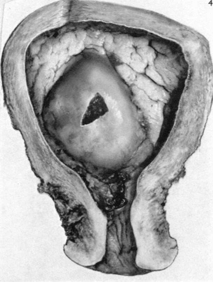
Fig. 4.
Photograph ( x 1-1) of a general view of the interior of the uterus containing the implantation site of a 10 mm. embryo (CX. 100).
A small opening has been made through the central part of the decidua capsularis.
Note the smoothness of this portion of the decidua in comparison with the furrowed decidua vera (parietalis).
Plates: 1 | 2 | 3 | 4 | 5 | 6 | 7 | 8 | 9 | 10 | 11 | 12 | 13
Plate 1
Plate 2
Plate 3
Plate 4
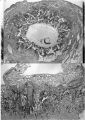
Plate 5
Plate 6
Plate 7
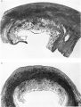
Plate 8
Plate 9
Plate 10
Plate 11
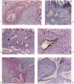
Plate 12
Plate 13


Reference
Hamilton WJ. and Boyd JD. Development of the human placenta in the first three months of gestation. (1960) J Anat. 94(3): 297-328. PMID14399291 | PDF
Cite this page: Hill, M.A. (2026, Mayıs 12) Embryology HamiltonBoyd1960 fig04.jpg. Retrieved from https://embryology.med.unsw.edu.au/embryology/index.php/File:HamiltonBoyd1960_fig04.jpg
- © Dr Mark Hill 2026, UNSW Embryology ISBN: 978 0 7334 2609 4 - UNSW CRICOS Provider Code No. 00098G
File history
Yi efo/eka'e gwa ebo wo le nyangagi wuncin ye kamina wunga tinya nan
| Gwalagizhi | Nyangagi | Dimensions | User | Comment | |
|---|---|---|---|---|---|
| current | 13:16, 6 August 2020 | | 700 × 926 (131 KB) | Z8600021 (talk | contribs) |
You cannot overwrite this file.
File usage
The following 2 pages use this file: