File:Early Development of Heart Tube.jpg
From Embryology
Size of this preview: 800 × 596 pixels. Other resolution: 1,475 × 1,099 pixels.
Original file (1,475 × 1,099 pixels, file size: 132 KB, MIME type: image/jpeg)
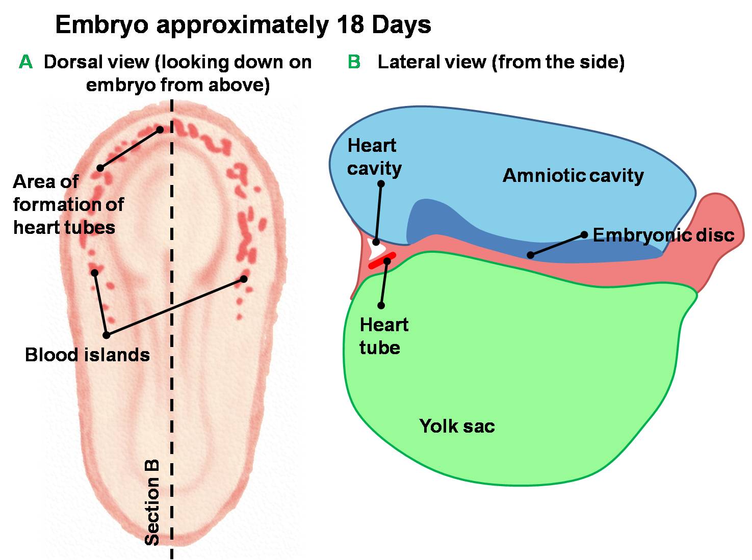
Early Development of Heart Tube
Dorsal and lateral views of the earliest stages of cardiac development in the human embryo.
Angiogenesis creates blood islands throughout the embryo during the third week of development.
Angioblastic cords form in the cardiogenic mesoderm and canalise to form the early heart tubes.

| Begin Basic | Primitive Heart Tube | Embryonic Heart Divisions | Vascular Heart Connections |
Cite this page: Hill, M.A. (2026, October 12) Embryology Early Development of Heart Tube.jpg. Retrieved from https://embryology.med.unsw.edu.au/embryology/index.php/File:Early_Development_of_Heart_Tube.jpg
- © Dr Mark Hill 2026, UNSW Embryology ISBN: 978 0 7334 2609 4 - UNSW CRICOS Provider Code No. 00098G
File history
Yi efo/eka'e gwa ebo wo le nyangagi wuncin ye kamina wunga tinya nan
| Gwalagizhi | Nyangagi | Dimensions | User | Comment | |
|---|---|---|---|---|---|
| current | 11:03, 14 March 2010 | | 1,475 × 1,099 (132 KB) | Z3212774 (talk | contribs) | Dorsal and lateral views of the earliest stages of cardiac development in the human embryo. Angiogenesis creates blood islands throughout the embryo during the third week of development. Angioblastic cords form in the cardiogenic mesoderm and canalise to |
You cannot overwrite this file.
File usage
The following 4 pages use this file:


