File:CameronMilligan1910 fig11.jpg

CameronMilligan1910_fig11.jpg (800 × 551 pixels, file size: 17 KB, MIME type: image/jpeg)
Fig. 11. Diagram to show the relation of the auditory syncytium to the hind brain and otic vesicle in an early vertebrate embryo
From a further examination of embryos of Cyclopterus lumpus it was ascertained that these subsidiary sense-epithelium patches were all derivatives of the originally single area, with which the auditory syncytium was in continuity during the early developmental stages (fig. 11).
Reference
Cameron J. and Milligan W. The development of the auditory nerve in vertebrates. (1910) J Anat. Physiol. 44(2): 111-32. PubMed 17232833
Cite this page: Hill, M.A. (2026, October 1) Embryology CameronMilligan1910 fig11.jpg. Retrieved from https://embryology.med.unsw.edu.au/embryology/index.php/File:CameronMilligan1910_fig11.jpg
- © Dr Mark Hill 2026, UNSW Embryology ISBN: 978 0 7334 2609 4 - UNSW CRICOS Provider Code No. 00098G
File history
Yi efo/eka'e gwa ebo wo le nyangagi wuncin ye kamina wunga tinya nan
| Gwalagizhi | Nyangagi | Dimensions | User | Comment | |
|---|---|---|---|---|---|
| current | 17:11, 6 November 2017 | | 800 × 551 (17 KB) | Z8600021 (talk | contribs) | |
| 17:10, 6 November 2017 | 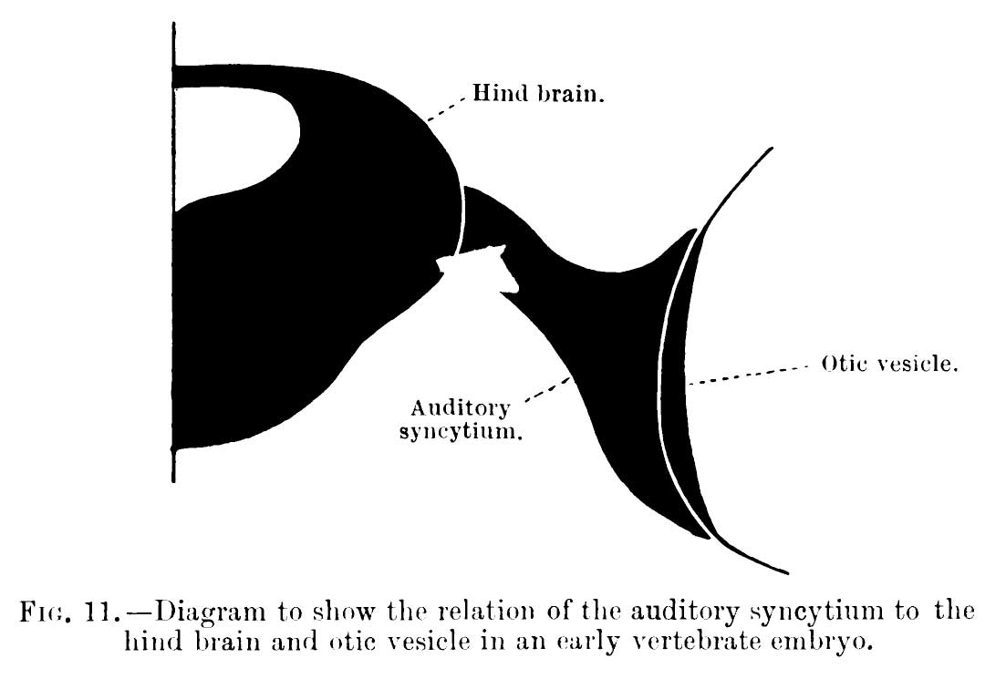 | 1,103 × 745 (39 KB) | Z8600021 (talk | contribs) |
You cannot overwrite this file.
File usage
The following page uses this file:


