File:Bowlby1882 fig03.jpg
From Embryology
Size of this preview: 522 × 599 pixels. Other resolution: 921 × 1,057 pixels.
Original file (921 × 1,057 pixels, file size: 256 KB, MIME type: image/jpeg)
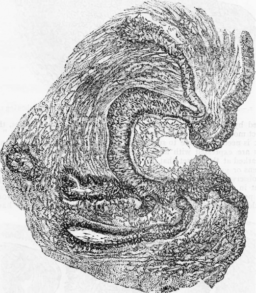
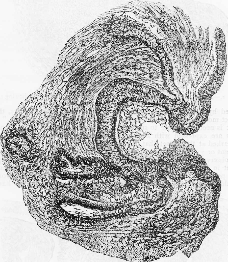
Fig. 3. Section of the mammary gland of a foetus aged seven months
The ingrowth is now hollowed, the bridge has broken away, and ducts are being developed from the epithelial ingrowth.
Reference
Bowlby AA. Development of the mammary gland. (1882) Br Med J. 2(1145): 1143-5. PMID 20750403
Cite this page: Hill, M.A. (2026, September 29) Embryology Bowlby1882 fig03.jpg. Retrieved from https://embryology.med.unsw.edu.au/embryology/index.php/File:Bowlby1882_fig03.jpg
- © Dr Mark Hill 2026, UNSW Embryology ISBN: 978 0 7334 2609 4 - UNSW CRICOS Provider Code No. 00098G
File history
Yi efo/eka'e gwa ebo wo le nyangagi wuncin ye kamina wunga tinya nan
| Gwalagizhi | Nyangagi | Dimensions | User | Comment | |
|---|---|---|---|---|---|
| current | 16:55, 13 August 2018 | | 921 × 1,057 (256 KB) | Z8600021 (talk | contribs) | |
| 16:54, 13 August 2018 | 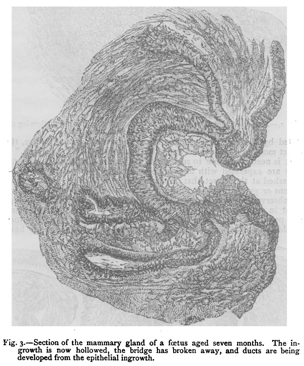 | 1,017 × 1,222 (189 KB) | Z8600021 (talk | contribs) | ==Fig. 3. Section of the mammary gland of a foetus aged seven months== The ingrowth is now hollowed, the bridge has broken away, and ducts are being developed from the epithelial ingrowth. |
You cannot overwrite this file.
File usage
The following page uses this file: