Category:Testis
From Embryology
This Embryology category shows media and pages related to the male gonad, the testis.
Pages in category 'Testis'
The following 86 pages are in this category, out of 86 total.
A
B
M
P
- Paper - A morphological study of testicular descent
- Paper - A study of the function of the epididymis 1 (1929)
- Paper - Development and transition of the testis, normal and abnormal 1
- Paper - Development and transition of the testis, normal and abnormal 2
- Paper - Development and transition of the testis, normal and abnormal 3
- Paper - Development and transition of the testis, normal and abnormal 4
- Paper - Development and vascularization of the testis (1906)
- Paper - Has a persistence of the Müllerian ducts any relation to the conditions of cryptorchidism?
- Paper - Studies on the fine structure of the mammalian testis 1
- Paper - The Embryonic Development of the Interstitial Cells of Leydig (1904)
- Paper - The histology of the retained testis in the human subject at different ages, and its comparison with the scrotal testis (1929)
- Paper - The inguinal canal in the foetus and new-born (1944)
- Paper - The morphology of the seminiferous tubules of Mammalia (1913)
- Template:Peritubular myoid cell
- Template:Persistent Müllerian duct syndrome
- Template:Primary spermatocyte
R
- Template:Ref-Allen1904
- Template:Ref-BascomOsterud1925
- Template:Ref-Bremer1911
- Template:Ref-BurgosFawcett1955
- Template:Ref-Cooper1929
- Template:Ref-Crew1922
- Template:Ref-Hill1906
- Template:Ref-Lockwood1887a
- Template:Ref-Lockwood1887b
- Template:Ref-Lockwood1888a
- Template:Ref-Lockwood1888b
- Template:Ref-PelliniemiNiemi1969
- Template:Ref-RowlandsBrambell1932
- Template:Ref-Young1929
- Template:Ref-Young1961
S
T
Media in category 'Testis'
The following 169 files are in this category, out of 169 total.
 Adrenal and gonad early development.jpg 700 × 397; 50 KB
 Adrenal and gonad steroidogenic factor 1 expression.jpg 1,000 × 636; 88 KB
 Bailey309.jpg 594 × 592; 58 KB
 Bailey327.jpg 872 × 567; 89 KB
 Bailey332.jpg 637 × 356; 53 KB
 Bailey337.jpg 790 × 573; 74 KB
 Bailey338.jpg 940 × 473; 87 KB
 Bailey341.jpg 832 × 675; 69 KB
 BurgosFawcett1955 fig11.jpg 1,453 × 2,015; 528 KB
 BurgosFawcett1955 fig13.jpg 1,460 × 2,049; 501 KB
 BurgosFawcett1955 fig14.jpg 1,456 × 1,965; 381 KB
 BurgosFawcett1955 text-fig01.jpg 1,280 × 1,137; 143 KB
 Corner1920 fig01.jpg 1,000 × 606; 159 KB
 Cryptorchidism.jpg 600 × 390; 35 KB
 Ductus deferens 01.jpg 400 × 533; 76 KB
 Ductus deferens 02.jpg 400 × 533; 80 KB
 Enrico Sertoli.jpg 650 × 800; 68 KB
 Epididymis histology 01.jpg 600 × 375; 20 KB
 Epididymis histology 02.jpg 400 × 534; 71 KB
 Epididymis histology 03.jpg 400 × 533; 68 KB
 Fetal gonad retinoid receptor expression 01.jpg 1,004 × 1,000; 226 KB
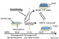 Gadd45g and sex determination model.jpg 909 × 600; 62 KB
 Germ cell tumor 02.jpg 800 × 599; 168 KB
 Gray1114.jpg 450 × 471; 47 KB
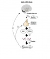 HPG male axis.jpg 600 × 700; 36 KB
 Human fetal gonad retinoid receptor expression.jpg 1,004 × 1,000; 447 KB
 Keibel Mall 008.jpg 1,200 × 515; 161 KB
 Keibel Mall 2 633.jpg 1,000 × 681; 74 KB
 Keibel Mall 2 635.jpg 1,000 × 1,073; 223 KB
 Keibel Mall 2 636.jpg 1,200 × 738; 119 KB
 Keibel Mall 2 658a.jpg 1,127 × 1,200; 103 KB
 Keibel Mall 2 658b.jpg 895 × 1,200; 98 KB
 Keith1902 fig102.jpg 800 × 605; 68 KB
 Keith1902 fig104.jpg 800 × 601; 77 KB
 Kollmann013.jpg 732 × 626; 95 KB
 Kollmann445.jpg 776 × 738; 112 KB
 Kollmann446.jpg 723 × 414; 38 KB
 Kollmann447.jpg 564 × 556; 42 KB
 Kollmann448.jpg 643 × 576; 43 KB
 Kollmann449.jpg 613 × 617; 44 KB
 Kollmann458.jpg 1,000 × 520; 120 KB
 Leydig cell PMID13693345 EM02.jpg 1,359 × 957; 341 KB
 Leydig cell PMID13693345 EM03.jpg 1,359 × 957; 325 KB
 Leydig cells stained for LHCGR1.jpg 404 × 322; 16 KB
 Lockwood1887b fig23.jpg 500 × 423; 41 KB
 Lockwood1887b fig24.jpg 600 × 463; 82 KB
 Lockwood1887b fig25.jpg 715 × 1,000; 125 KB
 Lockwood1887b fig26.jpg 500 × 266; 38 KB
 Lockwood1887b fig27.jpg 500 × 378; 46 KB
 Lockwood1887b fig28.jpg 600 × 592; 43 KB
 Lockwood1887b fig29.jpg 800 × 656; 121 KB
 Lockwood1887b fig30.jpg 800 × 619; 69 KB
 Lockwood1887b fig31.jpg 800 × 576; 49 KB
 Lockwood1887b fig32.jpg 500 × 329; 55 KB
 Lockwood1887b fig33.jpg 600 × 617; 101 KB
 Lockwood1887b fig34.jpg 408 × 800; 43 KB
 Lockwood1887b fig35.jpg 800 × 600; 117 KB
 Lockwood1887b fig36.jpg 600 × 828; 53 KB
 Lockwood1887b fig37.jpg 800 × 739; 102 KB
 Lockwood1887b fig38.jpg 800 × 688; 65 KB
 Lockwood1887b fig39.jpg 800 × 1,102; 93 KB
 Lockwood1887b fig40.jpg 774 × 1,000; 92 KB
 Lockwood1887b fig41.jpg 800 × 547; 129 KB
 Lockwood1887b plate02.jpg 2,998 × 2,272; 1.21 MB
 Lockwood1888a fig47.jpg 1,000 × 712; 279 KB
 Lockwood1888a fig50.jpg 959 × 652; 85 KB
 Lockwood1888a plate07.jpg 1,280 × 985; 285 KB
 Lockwood1888b fig49.jpg 800 × 496; 69 KB
 Lockwood1888b fig51.jpg 800 × 725; 88 KB
 Lockwood1888b fig52.jpg 800 × 824; 60 KB
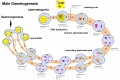 Male gametogenesis.jpg 1,000 × 666; 121 KB
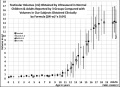 Male puberty testicular volume graph.jpg 1,140 × 826; 126 KB
 Model male androsterone synthesis.jpg 740 × 518; 92 KB
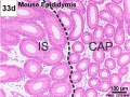
 Mouse epididymis development 01.jpg 1,200 × 909; 486 KB
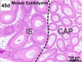
 Mouse epididymis development 02.jpg 600 × 451; 138 KB
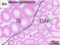
 Mouse epididymis development 03.jpg 600 × 451; 119 KB
 Mouse epididymis development 04.jpg 600 × 451; 120 KB
 Mouse epididymis development 05.jpg 600 × 451; 112 KB
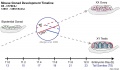 Mouse gonad development timeline.jpg 1,200 × 697; 98 KB
 Mouse gonad Gcnf expression 01.jpg 1,947 × 843; 304 KB
 Mouse gonad Gcnf expression E12.5.jpg 331 × 785; 68 KB
 Mouse gonad Gcnf expression E13.5.jpg 332 × 784; 60 KB
 Mouse gonad Gcnf expression E14.5.jpg 334 × 784; 60 KB
 Mouse gonad Gcnf expression E15.5.jpg 338 × 782; 53 KB
 Mouse gonad Gcnf expression E16.5.jpg 325 × 786; 40 KB
 Mouse gonad Gcnf expression E17.5.jpg 328 × 786; 44 KB
 Mouse gonad sex determination 01.jpg 600 × 600; 81 KB
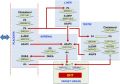
 Mouse sex determination genes 01.jpg 1,280 × 923; 73 KB
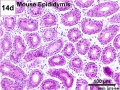
 Mouse- epididymis histology.jpg 751 × 383; 82 KB
 Mouse- gonadal supporting cell development.jpg 1,000 × 588; 74 KB
 Nelsen1953 fig002.jpg 1,200 × 1,037; 249 KB
 Nelsen1953 fig004.jpg 1,200 × 960; 200 KB
 Nelsen1953 fig005.jpg 1,200 × 709; 102 KB
 Nelsen1953 fig006.jpg 1,200 × 732; 224 KB
 Nelsen1953 fig007.jpg 1,200 × 1,311; 343 KB
 Orchidometer.jpg 361 × 225; 14 KB
 Rat blood–testis barrier 01.jpg 1,002 × 1,599; 221 KB
 Rat blood–testis barrier 02.jpg 1,002 × 853; 125 KB
 Rat- immortal germ cells are spermatogonial stem cells.jpg 459 × 1,000; 72 KB
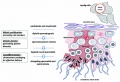 Seminiferous tubule cartoon.jpg 800 × 544; 92 KB
 Seminiferous-tubule-HEx40.jpg 400 × 500; 59 KB
 Simkins1928 plate01.jpg 1,574 × 2,003; 237 KB
 Simkins1928 plate02.jpg 1,464 × 2,126; 223 KB
 Simkins1928 plate03.jpg 1,551 × 2,086; 293 KB
 Simkins1928 plate04.jpg 1,543 × 2,026; 219 KB
 Simkins1928 plate05.jpg 1,281 × 2,111; 154 KB
 Simkins1928 plate06.jpg 1,587 × 2,088; 277 KB
 Simkins1928 plate07.jpg 1,540 × 2,096; 239 KB
 Simkins1928 plate08.jpg 1,558 × 1,797; 220 KB
 Simkins1928 plate09.jpg 1,548 × 2,096; 263 KB
 Simkins1928 plate10.jpg 1,565 × 1,386; 140 KB
 Spermatogenesis cartoon 01.jpg 1,064 × 759; 142 KB
 Spermatozoa histology 001.jpg 1,280 × 1,024; 366 KB
 Spermatozoa histology 002.jpg 1,280 × 1,024; 246 KB
 Spermatozoa histology 003.jpg 1,280 × 1,024; 166 KB
 Stage 22 image 188.jpg 1,000 × 665; 112 KB
 Stage 22 image 191.jpg 1,000 × 653; 100 KB
 Stage 22 image 194.jpg 1,000 × 671; 210 KB
 Stage 22 image 195.jpg 1,000 × 657; 265 KB
 Stage 22 image 201.jpg 1,200 × 754; 324 KB
 Stage 22 image 202.jpg 1,455 × 920; 617 KB
 Stage 22 image 301.jpg 1,200 × 754; 329 KB
 Stage 22 image 302.jpg 1,455 × 920; 625 KB
 Suprascrotal testis.jpg 1,000 × 751; 126 KB
 Testicular volume graph.jpg 554 × 405; 57 KB
 Testis histology 001.jpg 1,280 × 1,024; 574 KB
 Testis histology 002.jpg 1,280 × 1,024; 599 KB
 Testis histology 003.jpg 1,280 × 1,024; 183 KB
 Testis histology 004.jpg 1,280 × 1,024; 396 KB
 Testis histology 005.jpg 1,280 × 1,024; 266 KB
 Testis histology 006.jpg 1,280 × 1,024; 251 KB
 Testis histology 007.jpg 1,280 × 1,024; 256 KB
 Testis histology 008.jpg 1,280 × 1,024; 454 KB
 Testis histology 009.jpg 1,280 × 1,024; 339 KB
 Testis histology 010.jpg 1,280 × 1,024; 422 KB
 Testis histology 011.jpg 1,280 × 1,024; 245 KB
 Testis histology 012.jpg 1,280 × 1,024; 266 KB
 Testis histology 013.jpg 1,280 × 1,024; 418 KB
 Testis histology 014.jpg 1,280 × 1,024; 352 KB
 Testis histology 015.jpg 1,280 × 1,024; 281 KB
 Testis histology 016.jpg 1,280 × 1,024; 322 KB
 Testis histology 017.jpg 1,280 × 1,024; 283 KB
 Testis histology 018.jpg 1,280 × 1,024; 350 KB
 Testis histology 019.jpg 1,280 × 1,024; 239 KB
 Testis histology 02.jpg 246 × 481; 49 KB
 Testis histology 020.jpg 1,300 × 685; 334 KB
 Testis histology 021.jpg 1,200 × 962; 312 KB
 Testis histology 022.jpg 1,229 × 966; 311 KB
 Testis histology 023.jpg 600 × 375; 35 KB
 Testis histology 1.jpg 400 × 500; 113 KB
 Testis histology 2.jpg 400 × 500; 32 KB
 Testis histology.jpg 400 × 500; 54 KB
 Testis, young H&E reproductive system, male, convoluted seminiferous tubules x10.jpg 1,280 × 1,024; 396 KB
 Testis-descent end.jpg 600 × 480; 33 KB
 Testis-descent start.jpg 600 × 480; 26 KB
 Wyndham1943 fig01.jpg 800 × 660; 70 KB
 Wyndham1943 fig02.jpg 733 × 768; 172 KB
 Wyndham1943 fig03.jpg 756 × 754; 189 KB
 Wyndham1943 fig04.jpg 742 × 751; 195 KB
 Wyndham1943 fig05.jpg 632 × 741; 120 KB
 Wyndham1943 fig06.jpg 641 × 814; 166 KB
 Wyndham1943 fig07.jpg 779 × 650; 127 KB
 Wyndham1943 fig08.jpg 886 × 638; 171 KB
 Wyndham1943 fig09.jpg 636 × 1,000; 198 KB
 Wyndham1943 fig10.jpg 605 × 1,000; 183 KB
 Wyndham1943 fig11.jpg 1,000 × 447; 96 KB
 Wyndham1943 plate01.jpg 2,004 × 2,469; 719 KB
 Wyndham1943 plate02.jpg 2,004 × 2,471; 990 KB
 Wyndham1943 plate03.jpg 2,004 × 2,464; 1.08 MB


