Category:Rabbit
From Embryology
This Embryology category shows pages and media related to rabbit development. This animal was also used as one of the historic models of vertebrate development.
- Links: Rabbit Development | Animal Development
Pages in category 'Rabbit'
The following 56 pages are in this category, out of 56 total.
P
- Paper - Abdominal pregnancy in animals with an account of a case of multiple ectopic gestation in a rabbit (1932)
- Paper - Development and histogenesis of the human pineal organ
- Paper - Mechanism of ovulation in the rabbit 2
- Paper - Models of the pancreas in embryos of the pig, rabbit, cat, and man (1908)
- Paper - Persistent thyroglossal duct in a rabbit (1934)
- Paper - Placentation in the rabbit
- Paper - The Comparative Behavior of Mammalian Eggs in Vivo and in Vitro
- Paper - The development and significance of the cell columns in the ventral horn of the cervical and upper thoracic spinal cord of the rabbit (1941)
- Paper - The development in vitro of young rabbit embryos
- Paper - The development of the anterior post-otic somites in the rabbit
- Paper - The development of the hypophysis cerebri of the rabbit
- Paper - The development of the veins in the limbs of rabbit embryos
- Paper - The embryonic development of the ovary and testis of the mammals (1904)
- Paper - The history of the prochordal plate in the rabbit
- Paper - The pharyngeal pouches and their derivatives in the mammalia
- Paper - The regular occurrence of intestinal diverticula in embryos of the pig, rabbit and man
- Template:Pincus1935 figures
R
- Template:Rabbit
- Rabbit Development
- Template:Rabbit Links
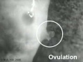
- Rabbit Ovulation Movie
- Template:Ref-Aasar1931
- Template:Ref-Atwell1918
- Template:Ref-Baxter1933
- Template:Ref-Bremer1912
- Template:Ref-Eales1932
- Template:Ref-Friedman1929
- Template:Ref-Hill1934
- Template:Ref-Houston1964
- Template:Ref-Hunter1934b
- Template:Ref-HunterRM1935
- Template:Ref-Lane-Claypon1905
- Template:Ref-Lewis1905a
- Template:Ref-Lewis1905b
- Template:Ref-LewisThyng1908
- Template:Ref-MinotTaylor1905
- Template:Ref-Murray1919
- Template:Ref-Pal1934
- Template:Ref-PMID17104355
- Template:Ref-PMID5969668
- Template:Ref-Romanes1941a
- Template:Ref-Thyng1908
- Template:Ref-Waddington1933a
- Template:Ref-WaddingtonWaterman1933
- Template:Ref-Wahl1915
Media in category 'Rabbit'
The following 183 files are in this category, out of 183 total.
 1hroldrabbit.jpg 800 × 532; 102 KB
 Aasar1931 plate1.jpg 1,160 × 2,287; 512 KB
 Aasar1931 plate2.jpg 1,185 × 2,258; 610 KB
 Aasar1931 plate3.jpg 1,507 × 2,284; 680 KB
 Aasar1931 plate4.jpg 1,512 × 2,271; 734 KB
 Aasar1931 text-fig01.jpg 715 × 956; 64 KB
 Allen1904 plate7.jpg 1,280 × 1,837; 704 KB
 Anson-1934 fig08-21.jpg 1,000 × 1,435; 178 KB
 Anson-1934 fig19.jpg 243 × 284; 11 KB
 Anson-1934 fig20.jpg 246 × 322; 9 KB
 Anson-1934 fig21.jpg 283 × 445; 16 KB
 Atwell1918 fig01.jpg 600 × 432; 61 KB
 Atwell1918 fig02.jpg 800 × 582; 83 KB
 Atwell1918 fig03.jpg 800 × 604; 117 KB
 Atwell1918 fig04.jpg 409 × 550; 37 KB
 Atwell1918 fig05.jpg 704 × 550; 56 KB
 Atwell1918 fig06.jpg 600 × 641; 84 KB
 Atwell1918 fig07.jpg 425 × 378; 44 KB
 Atwell1918 fig08.jpg 638 × 553; 46 KB
 Atwell1918 fig09.jpg 559 × 635; 84 KB
 Atwell1918 fig10.jpg 627 × 703; 90 KB
 Atwell1918 fig11.jpg 665 × 904; 131 KB
 Atwell1918 fig12.jpg 453 × 603; 36 KB
 Atwell1918 fig13.jpg 625 × 800; 122 KB
 Atwell1918 fig14.jpg 800 × 788; 117 KB
 Atwell1918 fig15.jpg 701 × 900; 171 KB
 Atwell1918 fig18.jpg 1,000 × 1,193; 168 KB
 Atwell1918 fig19.jpg 316 × 515; 21 KB
 Atwell1918 fig20.jpg 702 × 515; 48 KB
 Atwell1918 fig21.jpg 800 × 477; 108 KB
 Atwell1918 fig22.jpg 1,000 × 887; 100 KB
 Atwell1918 fig23.jpg 1,000 × 953; 123 KB
 Atwell1918 fig24.jpg 506 × 399; 43 KB
 Atwell1918 fig25.jpg 532 × 496; 48 KB
 Atwell1918 fig26.jpg 566 × 498; 57 KB
 Atwell1918 fig27.jpg 900 × 802; 113 KB
 Atwell1918 fig28.jpg 1,000 × 886; 348 KB
 Atwell1918 fig29.jpg 791 × 780; 120 KB
 Atwell1918 fig30.jpg 601 × 451; 45 KB
 Atwell1918 fig31.jpg 1,000 × 700; 267 KB
 Atwell1918 fig32.jpg 510 × 303; 28 KB
 Atwell1918 fig33.jpg 387 × 336; 29 KB
 Atwell1918 fig34.jpg 353 × 406; 32 KB
 Atwell1918 fig35.jpg 1,200 × 849; 386 KB
 Atwell1918 fig38.jpg 1,000 × 377; 129 KB
 Atwell1918 fig39.jpg 600 × 709; 139 KB
 Bailey078.jpg 706 × 321; 50 KB
 Bailey079.jpg 742 × 317; 51 KB
 Bailey161.jpg 645 × 629; 101 KB
 Bailey173.jpg 888 × 620; 113 KB
 Bailey203.jpg 406 × 614; 48 KB
 Bailey204.jpg 534 × 653; 62 KB
 Bailey205.jpg 534 × 653; 68 KB
 Bailey208.jpg 1,179 × 952; 165 KB
 Bailey209.jpg 1,248 × 988; 319 KB
 Bailey210.jpg 565 × 558; 63 KB
 Bailey211.jpg 767 × 820; 182 KB
 Bailey212.jpg 914 × 938; 218 KB
 Bailey215.jpg 944 × 958; 361 KB
 Bailey260.jpg 694 × 580; 58 KB
 Bailey291.jpg 868 × 313; 79 KB
 Bailey293.jpg 599 × 344; 32 KB
 Bailey296 297.jpg 513 × 726; 72 KB
 Bailey379-382.jpg 671 × 988; 199 KB
 Bailey465.jpg 806 × 931; 142 KB
 Bladder histology 004.jpg 1,280 × 1,024; 212 KB
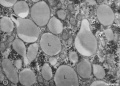
 Bone histology 017.jpg 1,280 × 1,024; 442 KB
 Bone histology 018.jpg 1,280 × 1,024; 336 KB
 Bone histology 019.jpg 1,280 × 1,024; 275 KB
 Bone histology 020.jpg 1,280 × 1,024; 272 KB
 Bone histology 021.jpg 1,280 × 1,024; 254 KB
 Early Growth of Trophoblast.pdf ; 234 KB
 Fetal rabbit neuroepithelial body 01.jpg 793 × 1,200; 98 KB
 Foster095.jpg 916 × 543; 114 KB
 Foster096.jpg 735 × 735; 54 KB
 Foster097.jpg 972 × 238; 35 KB
 Foster098.jpg 912 × 327; 30 KB
 Foster099.jpg 488 × 943; 90 KB
 Foster100.jpg 444 × 509; 37 KB
 Foster102.jpg 945 × 458; 90 KB
 Foster103.jpg 896 × 905; 87 KB
 Foster104.jpg 963 × 244; 51 KB
 Foster106.jpg 1,006 × 1,074; 269 KB
 Foster107.jpg 632 × 867; 94 KB
 Foster108.jpg 839 × 692; 101 KB
 Foster116.jpg 715 × 703; 75 KB
 Foster120.jpg 798 × 729; 98 KB
 Foster123.jpg 830 × 745; 132 KB
 Foster128.jpg 998 × 859; 223 KB
 Fox1908 fig70.jpg 1,220 × 718; 83 KB
 Fox1908 fig71.jpg 1,332 × 814; 162 KB
 Fox1908 fig72.jpg 1,521 × 970; 152 KB
 Fox1908 fig73.jpg 1,269 × 1,133; 156 KB
 GladstoneWakeley1937 fig04.jpg 1,272 × 1,062; 255 KB
 GladstoneWakeley1937 fig08.jpg 1,356 × 1,121; 490 KB
 Gray0597.jpg 700 × 496; 80 KB
 Gray0866.jpg 624 × 500; 65 KB
 Gray1182.jpg 800 × 982; 119 KB
 Isidro Martinez.jpg 305 × 345; 84 KB
 Keibel Mall 330.jpg 860 × 349; 61 KB
 Keibel Mall 331.jpg 880 × 635; 127 KB
 Keibel1905 plate01.jpg 1,514 × 2,000; 590 KB
 Keibel1905 plate02.jpg 1,477 × 2,000; 335 KB
 Keibel1905 plate03.jpg 1,503 × 2,000; 572 KB
 Kellicott 180.jpg 1,007 × 800; 77 KB
 Kellicott 181.jpg 865 × 800; 154 KB
 Kollmann069.jpg 667 × 800; 76 KB
 Kollmann528.jpg 744 × 587; 91 KB
 Kollmann751.jpg 691 × 348; 34 KB
 LewisFT1902 plate01-fig1-3.jpg 1,000 × 1,729; 205 KB
 LewisFT1902 plate01-fig1.jpg 1,000 × 894; 111 KB
 LewisFT1902 plate01-fig2-4.jpg 1,000 × 2,054; 236 KB
 LewisFT1902 plate01-fig2.jpg 1,000 × 1,076; 125 KB
 LewisFT1902 plate01-fig3.jpg 1,000 × 823; 93 KB
 LewisFT1902 plate01-fig4.jpg 1,000 × 969; 112 KB
 LewisFT1902 plate02-fig5-7.jpg 1,000 × 1,723; 186 KB
 LewisFT1902 plate02-fig5.jpg 1,000 × 744; 85 KB
 LewisFT1902 plate02-fig6-8.jpg 1,000 × 1,769; 189 KB
 LewisFT1902 plate02-fig6.jpg 1,000 × 778; 88 KB
 LewisFT1902 plate02-fig7.jpg 1,000 × 757; 97 KB
 LewisFT1902 plate02-fig8.jpg 1,000 × 769; 98 KB
 Liver- Kupffer cell and reticular fibre.jpg 600 × 800; 49 KB
 Liver-reticular fibre.jpg 700 × 875; 77 KB
 Lutein cell glycogen granule em01.jpg 1,149 × 749; 169 KB
 Lutein cell lipid and glycogen em01.jpg 1,156 × 828; 149 KB
 Lutein cell lipid and glycogen em02.jpg 1,109 × 796; 227 KB
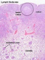
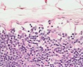 Lymph node 05.jpg 1,000 × 800; 180 KB
 Lymph node histology 02.jpg 450 × 600; 130 KB
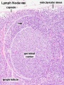 Lymph node histology 03.jpg 450 × 600; 140 KB
 Malformation1.jpg 354 × 570; 40 KB
 Minot1889 plate26.jpg 2,000 × 1,466; 587 KB
 Minot1889 plate28.jpg 2,000 × 1,257; 562 KB
 Minot1897 451.jpg 742 × 800; 145 KB
 Minot1897 fig004.jpg 633 × 543; 72 KB
 Minot1897 fig008.jpg 628 × 552; 118 KB
 Minot1897 fig086.jpg 1,020 × 948; 190 KB
 Minot1897 fig087.jpg 1,658 × 1,108; 457 KB
 Ovary Graaf.jpg 526 × 344; 83 KB
 Ovulation 001.mov ; 376 KB
- Ovulation 002.mov ; 422 KB
 Pincus1935-fig01.jpg 563 × 700; 101 KB
 Pincus1935-fig02.jpg 563 × 700; 60 KB
 Pincus1935-fig05.jpg 563 × 700; 82 KB
 Pincus1935-fig06.jpg 563 × 700; 69 KB
 Pincus1935-fig09.jpg 563 × 700; 66 KB
 Pincus1935-fig10.jpg 563 × 700; 61 KB
 Pincus1935-plate01.jpg 1,300 × 2,326; 463 KB
 Pincus1935-plate02.jpg 1,200 × 2,289; 446 KB
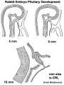 Pituitary rabbit development.jpg 374 × 500; 33 KB
 Pouchet1847 plate03.jpg 1,721 × 2,000; 456 KB
 Pouchet1847 plate04.jpg 1,787 × 2,000; 388 KB
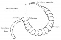 Rabbit caecum-appendix.jpg 1,000 × 655; 62 KB
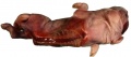
 Rabbit clone.jpg 672 × 224; 39 KB
 Rabbit gonad timeline.jpg 1,200 × 352; 71 KB
 Rabbit knee.jpg 669 × 575; 154 KB
 Rabbit-ovulation.jpg 320 × 240; 6 KB
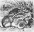
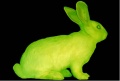
 Rabbit.jpg 278 × 156; 3 KB
 Rabbithhdrocephalus.jpg 795 × 485; 73 KB
 Rabbitmalformation3.jpg 363 × 574; 44 KB
 Rabbits.png 400 × 375; 133 KB
 Rabbitspinabifida1.jpg 819 × 360; 46 KB
 Stomach histology 007.jpg 1,280 × 1,024; 308 KB
 Testis histology 018.jpg 1,280 × 1,024; 350 KB
 Testis histology 019.jpg 1,280 × 1,024; 239 KB
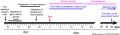
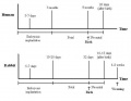 Timeline rabbit.JPG 633 × 488; 28 KB
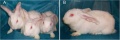
 Transgenic rabbit.jpg 453 × 306; 28 KB
 Williams1908-fig18.jpg 921 × 1,500; 89 KB
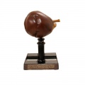
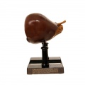
 Ziegler model 32.jpg 800 × 800; 41 KB
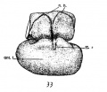
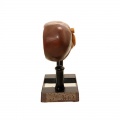
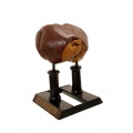
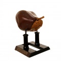
 Ziegler model 33.jpg 800 × 800; 28 KB
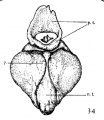
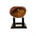
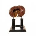
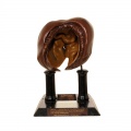
 Ziegler model 34.jpg 800 × 800; 34 KB
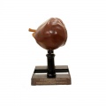
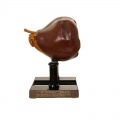
 Ziegler model 35.jpg 800 × 800; 29 KB
 Ziegler model 36.jpg 800 × 800; 34 KB
 Ziegler model 41.jpg 800 × 800; 30 KB
 Ziegler model 42.jpg 800 × 800; 33 KB
 Ziegler model 43.jpg 800 × 800; 38 KB
 Ziegler model 44.jpg 800 × 800; 43 KB
 Ziegler model 45.jpg 800 × 800; 33 KB
 Ziegler model 46.jpg 800 × 799; 35 KB
 Ziegler model 47.jpg 800 × 800; 40 KB
 Ziegler model 48.jpg 800 × 800; 46 KB
 Ziegler model legend 33.jpg 800 × 800; 74 KB
 Ziegler model legend 41.jpg 800 × 800; 64 KB
 Ziegler model legend 45.jpg 800 × 799; 0 bytes


