Category:Rabbit: Difference between revisions
From Embryology
mNo edit summary |
mNo edit summary |
||
| (4 intermediate revisions by the same user not shown) | |||
| Line 1: | Line 1: | ||
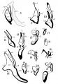
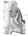
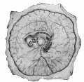
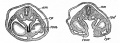
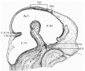
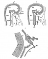
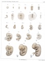
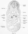
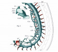
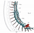
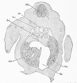
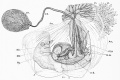
[[File:Keibel1905 plate03.jpg|thumb|link=Rabbit Development|1905 Rabbit Development]] | |||
This {{Embryology}} category shows pages and media related to rabbit development. The rabbit (Taxon- ''Oryctolagus cuniculus'') has been also used as one of the extensively used historic models of vertebrate development. Easy to breed, short development time course, and large litter sizes. First staged in 1905. | |||
{{Rabbit Links}} | |||
[[Category:Animal Development]] [[Category:Rabbit]] | [[Category:Animal Development]] [[Category:Rabbit]] | ||
Latest revision as of 12:30, 13 February 2017
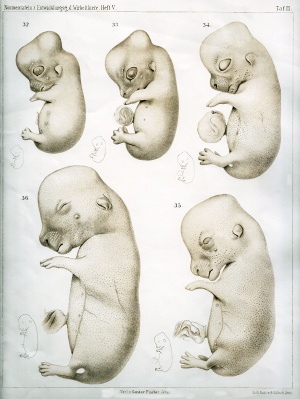
This Embryology category shows pages and media related to rabbit development. The rabbit (Taxon- Oryctolagus cuniculus) has been also used as one of the extensively used historic models of vertebrate development. Easy to breed, short development time course, and large litter sizes. First staged in 1905.
Pages in category 'Rabbit'
The following 56 pages are in this category, out of 56 total.
P
- Paper - Abdominal pregnancy in animals with an account of a case of multiple ectopic gestation in a rabbit (1932)
- Paper - Development and histogenesis of the human pineal organ
- Paper - Mechanism of ovulation in the rabbit 2
- Paper - Models of the pancreas in embryos of the pig, rabbit, cat, and man (1908)
- Paper - Persistent thyroglossal duct in a rabbit (1934)
- Paper - Placentation in the rabbit
- Paper - The Comparative Behavior of Mammalian Eggs in Vivo and in Vitro
- Paper - The development and significance of the cell columns in the ventral horn of the cervical and upper thoracic spinal cord of the rabbit (1941)
- Paper - The development in vitro of young rabbit embryos
- Paper - The development of the anterior post-otic somites in the rabbit
- Paper - The development of the hypophysis cerebri of the rabbit
- Paper - The development of the veins in the limbs of rabbit embryos
- Paper - The embryonic development of the ovary and testis of the mammals (1904)
- Paper - The history of the prochordal plate in the rabbit
- Paper - The pharyngeal pouches and their derivatives in the mammalia
- Paper - The regular occurrence of intestinal diverticula in embryos of the pig, rabbit and man
- Template:Pincus1935 figures
R
- Template:Rabbit
- Rabbit Development
- Template:Rabbit Links
- Rabbit Ovulation Movie
- Template:Ref-Aasar1931
- Template:Ref-Atwell1918
- Template:Ref-Baxter1933
- Template:Ref-Bremer1912
- Template:Ref-Eales1932
- Template:Ref-Friedman1929
- Template:Ref-Hill1934
- Template:Ref-Houston1964
- Template:Ref-Hunter1934b
- Template:Ref-HunterRM1935
- Template:Ref-Lane-Claypon1905
- Template:Ref-Lewis1905a
- Template:Ref-Lewis1905b
- Template:Ref-LewisThyng1908
- Template:Ref-MinotTaylor1905
- Template:Ref-Murray1919
- Template:Ref-Pal1934
- Template:Ref-PMID17104355
- Template:Ref-PMID5969668
- Template:Ref-Romanes1941a
- Template:Ref-Thyng1908
- Template:Ref-Waddington1933a
- Template:Ref-WaddingtonWaterman1933
- Template:Ref-Wahl1915
Media in category 'Rabbit'
The following 183 files are in this category, out of 183 total.
 1hroldrabbit.jpg 800 × 532; 102 KB
 Aasar1931 plate1.jpg 1,160 × 2,287; 512 KB
 Aasar1931 plate2.jpg 1,185 × 2,258; 610 KB
 Aasar1931 plate3.jpg 1,507 × 2,284; 680 KB
 Aasar1931 plate4.jpg 1,512 × 2,271; 734 KB
 Aasar1931 text-fig01.jpg 715 × 956; 64 KB
 Allen1904 plate7.jpg 1,280 × 1,837; 704 KB
 Anson-1934 fig08-21.jpg 1,000 × 1,435; 178 KB
 Anson-1934 fig19.jpg 243 × 284; 11 KB
 Anson-1934 fig20.jpg 246 × 322; 9 KB
 Anson-1934 fig21.jpg 283 × 445; 16 KB
 Atwell1918 fig01.jpg 600 × 432; 61 KB
 Atwell1918 fig02.jpg 800 × 582; 83 KB
 Atwell1918 fig03.jpg 800 × 604; 117 KB
 Atwell1918 fig04.jpg 409 × 550; 37 KB
 Atwell1918 fig05.jpg 704 × 550; 56 KB
 Atwell1918 fig06.jpg 600 × 641; 84 KB
 Atwell1918 fig07.jpg 425 × 378; 44 KB
 Atwell1918 fig08.jpg 638 × 553; 46 KB
 Atwell1918 fig09.jpg 559 × 635; 84 KB
 Atwell1918 fig10.jpg 627 × 703; 90 KB
 Atwell1918 fig11.jpg 665 × 904; 131 KB
 Atwell1918 fig12.jpg 453 × 603; 36 KB
 Atwell1918 fig13.jpg 625 × 800; 122 KB
 Atwell1918 fig14.jpg 800 × 788; 117 KB
 Atwell1918 fig15.jpg 701 × 900; 171 KB
 Atwell1918 fig18.jpg 1,000 × 1,193; 168 KB
 Atwell1918 fig19.jpg 316 × 515; 21 KB
 Atwell1918 fig20.jpg 702 × 515; 48 KB
 Atwell1918 fig21.jpg 800 × 477; 108 KB
 Atwell1918 fig22.jpg 1,000 × 887; 100 KB
 Atwell1918 fig23.jpg 1,000 × 953; 123 KB
 Atwell1918 fig24.jpg 506 × 399; 43 KB
 Atwell1918 fig25.jpg 532 × 496; 48 KB
 Atwell1918 fig26.jpg 566 × 498; 57 KB
 Atwell1918 fig27.jpg 900 × 802; 113 KB
 Atwell1918 fig28.jpg 1,000 × 886; 348 KB
 Atwell1918 fig29.jpg 791 × 780; 120 KB
 Atwell1918 fig30.jpg 601 × 451; 45 KB
 Atwell1918 fig31.jpg 1,000 × 700; 267 KB
 Atwell1918 fig32.jpg 510 × 303; 28 KB
 Atwell1918 fig33.jpg 387 × 336; 29 KB
 Atwell1918 fig34.jpg 353 × 406; 32 KB
 Atwell1918 fig35.jpg 1,200 × 849; 386 KB
 Atwell1918 fig38.jpg 1,000 × 377; 129 KB
 Atwell1918 fig39.jpg 600 × 709; 139 KB
 Bailey078.jpg 706 × 321; 50 KB
 Bailey079.jpg 742 × 317; 51 KB
 Bailey161.jpg 645 × 629; 101 KB
 Bailey173.jpg 888 × 620; 113 KB
 Bailey203.jpg 406 × 614; 48 KB
 Bailey204.jpg 534 × 653; 62 KB
 Bailey205.jpg 534 × 653; 68 KB
 Bailey208.jpg 1,179 × 952; 165 KB
 Bailey209.jpg 1,248 × 988; 319 KB
 Bailey210.jpg 565 × 558; 63 KB
 Bailey211.jpg 767 × 820; 182 KB
 Bailey212.jpg 914 × 938; 218 KB
 Bailey215.jpg 944 × 958; 361 KB
 Bailey260.jpg 694 × 580; 58 KB
 Bailey291.jpg 868 × 313; 79 KB
 Bailey293.jpg 599 × 344; 32 KB
 Bailey296 297.jpg 513 × 726; 72 KB
 Bailey379-382.jpg 671 × 988; 199 KB
 Bailey465.jpg 806 × 931; 142 KB
 Bladder histology 004.jpg 1,280 × 1,024; 212 KB
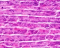
 Bone histology 017.jpg 1,280 × 1,024; 442 KB
 Bone histology 018.jpg 1,280 × 1,024; 336 KB
 Bone histology 019.jpg 1,280 × 1,024; 275 KB
 Bone histology 020.jpg 1,280 × 1,024; 272 KB
 Bone histology 021.jpg 1,280 × 1,024; 254 KB
 Early Growth of Trophoblast.pdf ; 234 KB
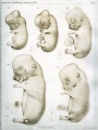
 Fetal rabbit neuroepithelial body 01.jpg 793 × 1,200; 98 KB
 Foster095.jpg 916 × 543; 114 KB
 Foster096.jpg 735 × 735; 54 KB
 Foster097.jpg 972 × 238; 35 KB
 Foster098.jpg 912 × 327; 30 KB
 Foster099.jpg 488 × 943; 90 KB
 Foster100.jpg 444 × 509; 37 KB
 Foster102.jpg 945 × 458; 90 KB
 Foster103.jpg 896 × 905; 87 KB
 Foster104.jpg 963 × 244; 51 KB
 Foster106.jpg 1,006 × 1,074; 269 KB
 Foster107.jpg 632 × 867; 94 KB
 Foster108.jpg 839 × 692; 101 KB
 Foster116.jpg 715 × 703; 75 KB
 Foster120.jpg 798 × 729; 98 KB
 Foster123.jpg 830 × 745; 132 KB
 Foster128.jpg 998 × 859; 223 KB
 Fox1908 fig70.jpg 1,220 × 718; 83 KB
 Fox1908 fig71.jpg 1,332 × 814; 162 KB
 Fox1908 fig72.jpg 1,521 × 970; 152 KB
 Fox1908 fig73.jpg 1,269 × 1,133; 156 KB
 GladstoneWakeley1937 fig04.jpg 1,272 × 1,062; 255 KB
 GladstoneWakeley1937 fig08.jpg 1,356 × 1,121; 490 KB
 Gray0597.jpg 700 × 496; 80 KB
 Gray0866.jpg 624 × 500; 65 KB
 Gray1182.jpg 800 × 982; 119 KB
 Isidro Martinez.jpg 305 × 345; 84 KB
 Keibel Mall 330.jpg 860 × 349; 61 KB
 Keibel Mall 331.jpg 880 × 635; 127 KB
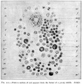
 Keibel1905 plate01.jpg 1,514 × 2,000; 590 KB
 Keibel1905 plate02.jpg 1,477 × 2,000; 335 KB
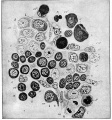
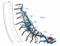
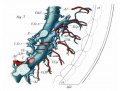
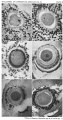
 Keibel1905 plate03.jpg 1,503 × 2,000; 572 KB
 Kellicott 180.jpg 1,007 × 800; 77 KB
 Kellicott 181.jpg 865 × 800; 154 KB
 Kollmann069.jpg 667 × 800; 76 KB
 Kollmann528.jpg 744 × 587; 91 KB
 Kollmann751.jpg 691 × 348; 34 KB
 LewisFT1902 plate01-fig1-3.jpg 1,000 × 1,729; 205 KB
 LewisFT1902 plate01-fig1.jpg 1,000 × 894; 111 KB
 LewisFT1902 plate01-fig2-4.jpg 1,000 × 2,054; 236 KB
 LewisFT1902 plate01-fig2.jpg 1,000 × 1,076; 125 KB
 LewisFT1902 plate01-fig3.jpg 1,000 × 823; 93 KB
 LewisFT1902 plate01-fig4.jpg 1,000 × 969; 112 KB
 LewisFT1902 plate02-fig5-7.jpg 1,000 × 1,723; 186 KB
 LewisFT1902 plate02-fig5.jpg 1,000 × 744; 85 KB
 LewisFT1902 plate02-fig6-8.jpg 1,000 × 1,769; 189 KB
 LewisFT1902 plate02-fig6.jpg 1,000 × 778; 88 KB
 LewisFT1902 plate02-fig7.jpg 1,000 × 757; 97 KB
 LewisFT1902 plate02-fig8.jpg 1,000 × 769; 98 KB
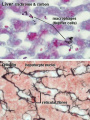 Liver- Kupffer cell and reticular fibre.jpg 600 × 800; 49 KB
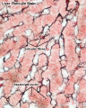 Liver-reticular fibre.jpg 700 × 875; 77 KB
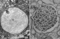 Lutein cell glycogen granule em01.jpg 1,149 × 749; 169 KB
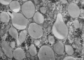 Lutein cell lipid and glycogen em01.jpg 1,156 × 828; 149 KB
 Lutein cell lipid and glycogen em02.jpg 1,109 × 796; 227 KB
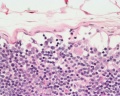 Lymph node 05.jpg 1,000 × 800; 180 KB
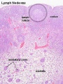 Lymph node histology 02.jpg 450 × 600; 130 KB
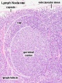 Lymph node histology 03.jpg 450 × 600; 140 KB
 Malformation1.jpg 354 × 570; 40 KB
 Minot1889 plate26.jpg 2,000 × 1,466; 587 KB
 Minot1889 plate28.jpg 2,000 × 1,257; 562 KB
 Minot1897 451.jpg 742 × 800; 145 KB
 Minot1897 fig004.jpg 633 × 543; 72 KB
 Minot1897 fig008.jpg 628 × 552; 118 KB
 Minot1897 fig086.jpg 1,020 × 948; 190 KB
 Minot1897 fig087.jpg 1,658 × 1,108; 457 KB
 Ovary Graaf.jpg 526 × 344; 83 KB
 Ovulation 001.mov ; 376 KB
- Ovulation 002.mov ; 422 KB
 Pincus1935-fig01.jpg 563 × 700; 101 KB
 Pincus1935-fig02.jpg 563 × 700; 60 KB
 Pincus1935-fig05.jpg 563 × 700; 82 KB
 Pincus1935-fig06.jpg 563 × 700; 69 KB
 Pincus1935-fig09.jpg 563 × 700; 66 KB
 Pincus1935-fig10.jpg 563 × 700; 61 KB
 Pincus1935-plate01.jpg 1,300 × 2,326; 463 KB
 Pincus1935-plate02.jpg 1,200 × 2,289; 446 KB
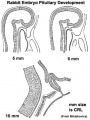 Pituitary rabbit development.jpg 374 × 500; 33 KB
 Pouchet1847 plate03.jpg 1,721 × 2,000; 456 KB
 Pouchet1847 plate04.jpg 1,787 × 2,000; 388 KB
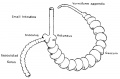 Rabbit caecum-appendix.jpg 1,000 × 655; 62 KB
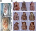
 Rabbit clone.jpg 672 × 224; 39 KB
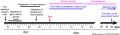 Rabbit gonad timeline.jpg 1,200 × 352; 71 KB
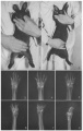
 Rabbit knee.jpg 669 × 575; 154 KB
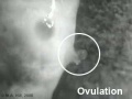 Rabbit-ovulation.jpg 320 × 240; 6 KB
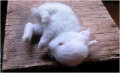
 Rabbit.jpg 278 × 156; 3 KB
 Rabbithhdrocephalus.jpg 795 × 485; 73 KB
 Rabbitmalformation3.jpg 363 × 574; 44 KB
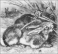 Rabbits.png 400 × 375; 133 KB
 Rabbitspinabifida1.jpg 819 × 360; 46 KB
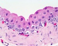
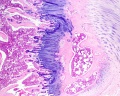
 Stomach histology 007.jpg 1,280 × 1,024; 308 KB
 Testis histology 018.jpg 1,280 × 1,024; 350 KB
 Testis histology 019.jpg 1,280 × 1,024; 239 KB
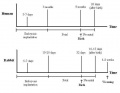 Timeline rabbit.JPG 633 × 488; 28 KB
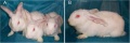
 Transgenic rabbit.jpg 453 × 306; 28 KB
 Williams1908-fig18.jpg 921 × 1,500; 89 KB
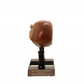
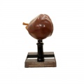
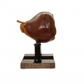
 Ziegler model 32.jpg 800 × 800; 41 KB
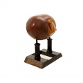
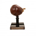
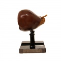
 Ziegler model 33.jpg 800 × 800; 28 KB
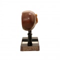
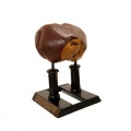
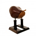
 Ziegler model 34.jpg 800 × 800; 34 KB
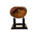
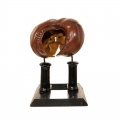
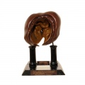
 Ziegler model 35.jpg 800 × 800; 29 KB
 Ziegler model 36.jpg 800 × 800; 34 KB
 Ziegler model 41.jpg 800 × 800; 30 KB
 Ziegler model 42.jpg 800 × 800; 33 KB
 Ziegler model 43.jpg 800 × 800; 38 KB
 Ziegler model 44.jpg 800 × 800; 43 KB
 Ziegler model 45.jpg 800 × 800; 33 KB
 Ziegler model 46.jpg 800 × 799; 35 KB
 Ziegler model 47.jpg 800 × 800; 40 KB
 Ziegler model 48.jpg 800 × 800; 46 KB
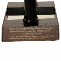 Ziegler model legend 33.jpg 800 × 800; 74 KB
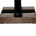 Ziegler model legend 41.jpg 800 × 800; 64 KB
 Ziegler model legend 45.jpg 800 × 799; 0 bytes


