Category:Palate
From Embryology
This Embryology category shows pages and media related to palate development and the associated abnormality of cleft palate/lip..
- Links: Palate Development
| Palate Links: palate | cleft lip and palate | cleft palate | head | Category:Palate |
Pages in category 'Palate'
The following 33 pages are in this category, out of 33 total.
A
B
C
P
R
Media in category 'Palate'
The following 93 files are in this category, out of 93 total.
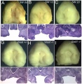
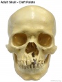 Adult skull cleft palate 01.jpg 750 × 1,000; 89 KB
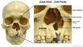 Adult skull cleft palate 02.jpg 1,280 × 720; 139 KB
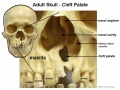 Adult skull cleft palate 03.jpg 904 × 678; 104 KB
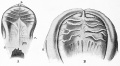
 Bailey140.jpg 793 × 505; 58 KB
 Bailey141.jpg 761 × 323; 66 KB
 Baumgartner1916 plate01.jpg 1,692 × 2,228; 308 KB
 Baumgartner1916 plate02.jpg 1,613 × 2,204; 399 KB
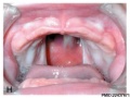
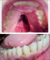
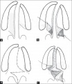
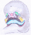
 Bilateral cleft palate.jpg 214 × 300; 11 KB
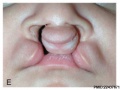
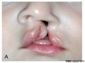 Cleft lip 001.jpg 400 × 300; 16 KB
 Cleft lip 002.jpg 400 × 300; 22 KB
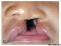 Cleft lip 003.jpg 400 × 300; 19 KB
 Cleft lip 004.jpg 400 × 300; 21 KB
 Cleft lip 005.jpg 400 × 300; 0 bytes
 Cleft lip 006.jpg 400 × 300; 22 KB
 Cleft lip 007.jpg 405 × 504; 38 KB
 Cleft lip 02.jpg 641 × 362; 22 KB
 Cleft lip 2.jpg 500 × 500; 160 KB
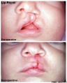
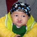
 Cleft lip.jpg 375 × 500; 101 KB
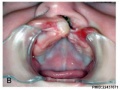
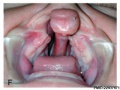
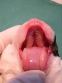
 Cleft palate 001.jpg 400 × 300; 22 KB
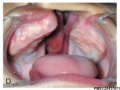
 Cleft palate 002.jpg 400 × 300; 20 KB
 Cleft palate 003.jpg 407 × 600; 46 KB
 Cleft palate 02.jpg 600 × 463; 34 KB
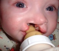
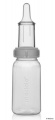 Cleft palate feeder.jpg 276 × 600; 19 KB
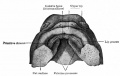
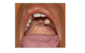
 Cleft Palate Maxillary View.jpg 498 × 599; 80 KB
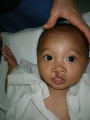
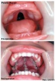
 Cleft palate.jpg 653 × 776; 124 KB
 Cleft Palate.jpg 698 × 600; 71 KB
 Dog day0-cleft palate.jpg 490 × 653; 39 KB
 Fawcett1906 01.jpg 760 × 780; 72 KB
 Fawcett1906 02.jpg 717 × 737; 58 KB
 Fawcett1906 03.jpg 706 × 700; 50 KB
 Fawcett1906 03a.jpg 581 × 750; 66 KB
 Fawcett1906 04.jpg 570 × 580; 22 KB
 Fawcett1906 05.jpg 633 × 700; 38 KB
 Fawcett1906 06.jpg 590 × 600; 26 KB
 Fawcett1906 07.jpg 563 × 603; 34 KB
 Fawcett1906 08.jpg 637 × 680; 38 KB
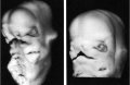
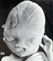
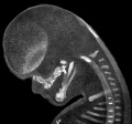
 Fetal head section 01.jpg 1,200 × 821; 186 KB
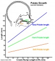 Fetal palate growth graph.jpg 681 × 757; 77 KB
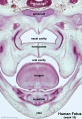
 Fetal week 10 hard palate 02.jpg 398 × 633; 66 KB
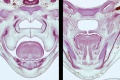 Fetal week 10 hard palate 04.jpg 1,198 × 795; 196 KB
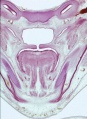
 Fetal week 10 hard palate 06.jpg 534 × 778; 88 KB
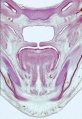
 Fetal week 10 hard palate 07.jpg 534 × 778; 97 KB
 Fetal week 10 palate 01.gif 534 × 778; 1.14 MB
 Fetal week 10 palate 01.mp4 ; 427 KB
 Fetal week 10 palate icon.jpg 534 × 778; 100 KB
 Fetal week 10 soft palate 01.jpg 571 × 784; 95 KB
 Fetal week 10 soft palate 02.jpg 534 × 778; 87 KB
 Fetal week 10 soft palate 03.jpg 534 × 778; 95 KB
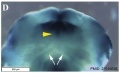
 Fetal week 9 hard palate fusion 01.jpg 661 × 400; 51 KB
 Fgf signalling in palate development.PNG 1,634 × 1,942; 2.33 MB
 File-Cleft palate in newborn mice.jpg 669 × 240; 26 KB
 Furlow Z-plasty technique.jpg 670 × 771; 147 KB
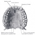
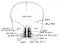
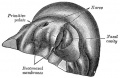 Gray0049.jpg 613 × 400; 61 KB
 Gray0050.jpg 600 × 332; 46 KB
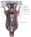
 Gray0859.jpg 694 × 600; 114 KB
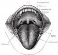
 Gray0996.jpg 800 × 822; 118 KB
 Gray1028.jpg 800 × 960; 202 KB
 Gray1201.jpg 581 × 550; 88 KB
 Human Embryo Stage18-19.jpg 600 × 392; 17 KB
 In vitro fetal palate explant culture.jpg 506 × 519; 213 KB
 Keibel Mall 2 256.jpg 1,543 × 850; 181 KB
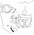
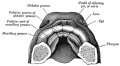
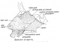 Keith1902 fig003.jpg 1,000 × 700; 113 KB
 Keith1902 fig007.jpg 946 × 700; 80 KB
 Keith1902 fig008.jpg 925 × 700; 122 KB
 Keith1902 fig009.jpg 1,000 × 568; 78 KB
 Keith1902 fig011.jpg 954 × 500; 66 KB
 Macklin-plate04.jpg 2,331 × 3,061; 956 KB
 Mall Meyer1921 fig86.jpg 666 × 762; 81 KB
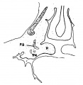
 Mall1906 fig04.jpg 1,114 × 1,044; 137 KB
 Mall1917 fig10.jpg 1,000 × 507; 62 KB
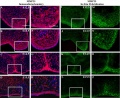 Mouse - palate MMP-25 expression.jpg 1,000 × 818; 243 KB
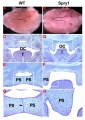 Mouse - Spry1 cleft palate.jpg 600 × 848; 166 KB
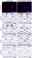
 Mouse E13.5 Bmp7 palate 1.jpg 1,200 × 993; 156 KB
 Mouse E13.5 Bmp7 palate 2.jpg 917 × 800; 88 KB
 Mouse E13.5 Bmp7 palate 3.jpg 800 × 488; 40 KB
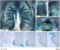
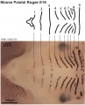 Mouse palatal rugae pattern E16.jpg 569 × 700; 56 KB
 Mouse palate increasing p63.jpg 654 × 1,199; 314 KB
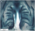
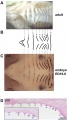 Mouse ruga pattern.jpg 600 × 1,059; 118 KB
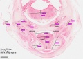
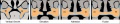 Normal palate shelf and key stages of mouse palatal development.jpg 771 × 153; 80 KB
 Palatal shelves animation.gif 550 × 400; 134 KB
 Palate and lip development animation.gif 536 × 600; 242 KB
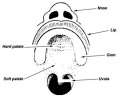
 Palate cartoon 01.jpg 700 × 539; 77 KB
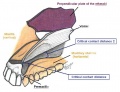
 Palate structure cartoon.jpg 300 × 242; 14 KB
 Repaired Cleft Palate.PNG 840 × 525; 473 KB
 Stage 22 image 219.jpg 1,250 × 892; 295 KB
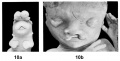
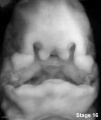 Stage16 cleft palate.jpg 337 × 400; 14 KB
 Stage16-18 face animation.gif 181 × 228; 80 KB
 Stage17 bf10.jpg 1,375 × 2,048; 134 KB
 Stage17 bf11.jpg 1,375 × 2,048; 166 KB
 Stage17-18 Primary palate.gif 458 × 283; 373 KB
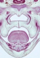
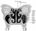
 Stage23 embryo oral cavity 03.jpg 1,000 × 693; 83 KB
 Stage23 embryo oral cavity 04.jpg 1,000 × 693; 90 KB
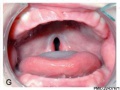
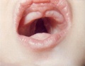
 Unilateral cleft palate.jpg 214 × 300; 12 KB


