Category:Ovary
From Embryology
The following media and pages relate to the ovary.
Subcategories
This category has the following 5 subcategories, out of 5 total.
Pages in category 'Ovary'
The following 133 pages are in this category, out of 133 total.
B
C
E
F
G
M
O
- Template:Oestrogen
- Template:Oestrogens
- Template:Oogenesis
- Template:Ova
- Template:Ovarian reserve
- Template:Ovary
- Template:Ovary abnormalities
- Ovary Development
- Ovary Development Movie
- Template:Ovary timeline
- Template:Ovary timeline table
- Template:Ovary Vignette
- Template:Ovulation
- Ovulation in the human ovary - Its mechanism and anomalies
- Ovulation Movie
P
- Paper - Cyclic changes in the ovaries and uterus of swine and their relations to the mechanism of implantation (1921)
- Paper - Development of the human ovary from birth to sexual maturity
- Paper - Human ova from large follicles
- Paper - Human ova from large follicles - including a search for maturation divisions and observations on atresia
- Paper - Human ova from large follicles - including a search for maturation divisions and observations on atresia (1930)
- Paper - Notes on the formation, structure and physiology of the corpus luteum of man, the pig, and the duck-billed platypus (1924)
- Paper - Selective elimination of ova in the adult ovary
- Paper - Selective elimination of ova in the adult ovary (1925)
- Paper - The corpus luteum in the ovary of the chicken (1918)
- Paper - The events of the primate ovarian cycle
- Paper - The fate of the graafian follicle in the human ovary
- Paper - The hormone of the corpus luteum
- Paper - The morphogenesis of the mammalian ovary (1913)
- Paper - The occurrence of polyovular graafian follicles (1924)
- Paper - The origin of the lutein cells of the corpus luteum
- Paper - The physiological descent of the ovaries in the human foetus
- Template:PCOS
- Template:Polycystic ovarian syndrome
- Template:Polycystic ovary syndrome
- Template:Preovulatory follicle
- Template:Primary follicle
- Template:Primordial follicle
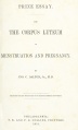
- Prize Essay on the Corpus Luteum (1851) 1
- Prize Essay on the Corpus Luteum (1851) 2
- Prize Essay on the Corpus Luteum (1851) 3
- Prize Essay on the Corpus Luteum (1851) Plates
- Template:Progesterone
R
- Rabbit Ovulation Movie
- Template:Ref-AdamsHertig1969a
- Template:Ref-AdamsHertig1969b
- Template:Ref-Allen1904
- Template:Ref-Allen1925
- Template:Ref-Allen1930
- Template:Ref-Allen1930b
- Template:Ref-Arai1920
- Template:Ref-Arnold1912
- Template:Ref-Brambell1927a
- Template:Ref-Brambell1928
- Template:Ref-Catchpole1949
- Template:Ref-Clark1899b
- Template:Ref-ClarkGJ1900
- Template:Ref-Corner1919
- Template:Ref-Corner1923
- Template:Ref-Corner1937
- Template:Ref-Corner1937b
- Template:Ref-Corner1952
- Template:Ref-Falkiner1933
- Template:Ref-Guttmacher1921
- Template:Ref-Hart1909d
- Template:Ref-Kampmeier1929
- Template:Ref-Kennedy1924
- Template:Ref-Kingsbury1913
- Template:Ref-Kingsbury1914
- Template:Ref-Kingsbury1914a
- Template:Ref-PearlBoring1918
- Template:Ref-Shaw1925
- Template:Ref-Shaw1926
- Template:Ref-Simkins1932
- Template:Ref-Solomons1924
- Template:Ref-Thomson1919a
- Template:Ref-Thomson1919b
- Template:Ref-White1951
- Template:Ref-Young1961
- Template:RU 486
- Template:RU486
S
Media in category 'Ovary'
The following 173 files are in this category, out of 173 total.
 Adrenal and gonad early development.jpg 700 × 397; 50 KB
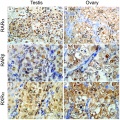
 Adrenal and gonad steroidogenic factor 1 expression.jpg 1,000 × 636; 88 KB
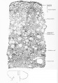
 Bailey001.jpg 850 × 794; 155 KB
 Bailey014.jpg 704 × 587; 116 KB
 Bailey328.jpg 704 × 795; 85 KB
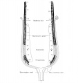
 Bailey329.jpg 838 × 381; 70 KB
 Bailey330.jpg 680 × 539; 109 KB
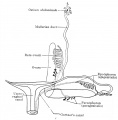
 Bailey331.jpg 890 × 782; 118 KB
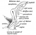
 Bailey335.jpg 590 × 468; 55 KB
 Bailey339.jpg 802 × 602; 111 KB
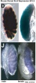
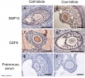 Bovine ovarian follicle BMP15 and GDF9 expression.jpg 874 × 800; 162 KB
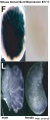
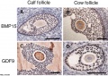 Bovine ovarian follicle BMP15 and GDF9.jpg 1,143 × 810; 219 KB
 Brambell1927a plate28.jpg 1,500 × 1,862; 169 KB
 Brambell1927a plate29.jpg 1,753 × 2,464; 290 KB
 Brambell1927a plate30.jpg 1,732 × 2,386; 451 KB
 Brambell1927a plate31.jpg 1,739 × 2,157; 444 KB
 Cat embryo ovary.jpg 505 × 492; 47 KB
 Corner-table02.jpg 800 × 647; 107 KB
 Corner001.jpg 858 × 356; 44 KB
 Corner002.jpg 873 × 723; 88 KB
 Corner002a.jpg 848 × 307; 49 KB
 Corner002b.jpg 849 × 328; 33 KB
 Corner1920 fig01.jpg 1,000 × 606; 159 KB
 Corner1920 fig02-05.jpg 1,000 × 671; 216 KB
 Corner1920 Plate 1.jpg 756 × 1,000; 222 KB
 Corpus luteum lutein cells.jpg 450 × 600; 104 KB
 Corpus luteum.jpg 450 × 600; 94 KB
 Dalton1851 fig01.jpg 538 × 800; 101 KB
 Dalton1851 fig02.jpg 717 × 800; 77 KB
 Dalton1851 fig03.jpg 681 × 800; 76 KB
 Dalton1851 fig04.jpg 734 × 800; 116 KB
 Dalton1851 fig05.jpg 706 × 800; 105 KB
 Dalton1851 fig06.jpg 599 × 800; 75 KB
 Dalton1851 plate01.jpg 719 × 1,200; 171 KB
 Dalton1851 plate02.jpg 721 × 1,200; 194 KB
 Dalton1851 titlepage.jpg 604 × 1,000; 49 KB
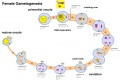 Female gametogenesis.jpg 1,000 × 666; 94 KB
 Fetal adrenal ectopic germ cells 01.jpg 1,092 × 1,280; 358 KB
 Fetal adrenal ectopic germ cells 02.jpg 1,086 × 446; 124 KB
 Fetal gonad retinoid receptor expression 01.jpg 1,004 × 1,000; 226 KB
 Fetal ovary meiosis 01.jpg 1,280 × 410; 132 KB
 Fetal ovary meiosis 02.jpg 496 × 600; 77 KB
 Fetal ovary meiosis 03.jpg 652 × 400; 64 KB
 Gray0589.jpg 900 × 534; 134 KB
 Gray1108.jpg 590 × 400; 73 KB
 Gray1112.jpg 550 × 548; 51 KB
 Gray1113.jpg 600 × 385; 68 KB
 Gray1161.jpg 1,000 × 671; 138 KB
 Gray1163.jpg 600 × 442; 91 KB
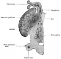
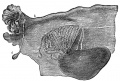
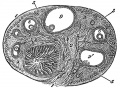
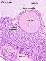
 Historic-ovary.jpg 385 × 283; 34 KB
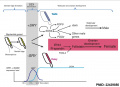
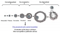
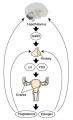
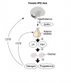 HPG female axis.jpg 600 × 700; 41 KB
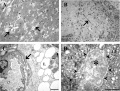 Human corpus luteum - light-and-electron-micrograph.jpg 936 × 711; 208 KB
 Human fetal gonad retinoid receptor expression.jpg 1,004 × 1,000; 447 KB
 Human fetal ovary FOXL2.jpg 739 × 1,087; 300 KB
 Human fetal ovary SMAD6 expression.jpg 711 × 535; 167 KB
 Human infant ovary follicle 01.jpg 800 × 800; 107 KB
 Human ovary - corpus luteum 01.jpg 1,024 × 979; 162 KB
 Human ovary - corpus luteum 02.jpg 837 × 800; 119 KB
 Human ovary - corpus luteum 11.jpg 1,024 × 979; 89 KB
 Human ovary - corpus luteum 21.jpg 1,024 × 979; 89 KB
 Human ovary follicle basement membrane 01.jpg 660 × 800; 184 KB
 Human ovary follicle basement membrane EM01.jpg 558 × 697; 100 KB
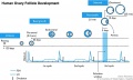 Human ovary follicle development.jpg 700 × 418; 50 KB
 Human ovary follicles light and electron microscopy 01.jpg 586 × 1,080; 225 KB
 Human ovary non-growing follicle model 02.jpg 1,200 × 918; 161 KB
 Human ovary non-growing follicle model.jpg 1,151 × 679; 116 KB
 Human ovary postnatal growth.jpg 800 × 467; 40 KB
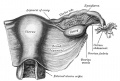
 Human ovulation 01.jpg 1,000 × 799; 175 KB
 Human ovulation 02.jpg 734 × 583; 68 KB
 Human ovulation 03.jpg 734 × 583; 76 KB
 Human ovulation 04.jpg 734 × 583; 64 KB
 Human ovulation 05.jpg 734 × 583; 71 KB
 Human ovulation 06.jpg 734 × 583; 82 KB
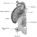
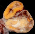
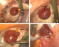
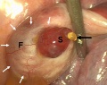
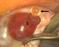
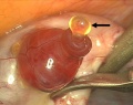
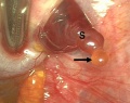
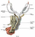
 Human right ovary and tube 1.jpg 916 × 680; 32 KB
 Human- adult ovary epithelial cords and primary follicles.jpg 600 × 384; 51 KB
 Human- adult ovary primary follicles.jpg 600 × 639; 56 KB
 Human- ovary age primary follicle numbers.jpg 600 × 520; 36 KB
 Infant ovary.jpg 943 × 571; 108 KB
 Keibel Mall 001.jpg 781 × 733; 138 KB
 Keibel Mall 008.jpg 1,200 × 515; 161 KB
 Keibel Mall 2 621.jpg 1,280 × 1,842; 589 KB
 Keibel Mall 2 658a.jpg 1,127 × 1,200; 103 KB
 Keibel Mall 2 658c.jpg 1,000 × 1,019; 90 KB
 Keith1902 fig081.jpg 818 × 800; 113 KB
 Kollmann012.jpg 696 × 437; 31 KB
 Kollmann451.jpg 796 × 826; 102 KB
 Kollmann452.jpg 687 × 606; 74 KB
 Kollmann461.jpg 721 × 528; 100 KB
 Kollmann462.jpg 758 × 429; 63 KB
 Lutein cell glycogen granule em01.jpg 1,149 × 749; 169 KB
 Lutein cell lipid and glycogen em01.jpg 1,156 × 828; 149 KB
 Lutein cell lipid and glycogen em02.jpg 1,109 × 796; 227 KB
 Luteoma of pregnancy 01.jpg 1,200 × 677; 177 KB
 Luteoma of pregnancy 02.jpg 1,200 × 831; 188 KB
 Model of human fetal ovarian cord development 01.jpg 800 × 330; 104 KB
 Monkey- ovary primordial follicle.jpg 1,000 × 800; 292 KB
 Mouse gonad development timeline.jpg 1,200 × 697; 98 KB
 Mouse gonad Gcnf expression 01.jpg 1,947 × 843; 304 KB
 Mouse gonad Gcnf expression E12.5.jpg 331 × 785; 68 KB
 Mouse gonad Gcnf expression E13.5.jpg 332 × 784; 60 KB
 Mouse gonad Gcnf expression E14.5.jpg 334 × 784; 60 KB
 Mouse gonad Gcnf expression E15.5.jpg 338 × 782; 53 KB
 Mouse gonad Gcnf expression E16.5.jpg 325 × 786; 40 KB
 Mouse gonad Gcnf expression E17.5.jpg 328 × 786; 44 KB
 Mouse model of ovarian cord formation 01.jpg 800 × 491; 85 KB
 Mouse model of ovarian cord formation.jpg 800 × 491; 85 KB
 Mouse newborn ovary day 1.mp4 ; 646 KB
- Mouse newborn ovary day 2-3.5.mp4 ; 1.95 MB
 Mouse oogenesis 01.jpg 1,781 × 1,222; 173 KB
 Mouse oogenesis 02.jpg 1,386 × 355; 40 KB
 Mouse ovarian follicle size.jpg 600 × 407; 36 KB
 Mouse ovary 01.jpg 602 × 482; 69 KB
 Mouse ovary normal and polycystic ovary syndrome 01.jpg 913 × 815; 356 KB
 Mouse sex determination genes 01.jpg 1,280 × 923; 73 KB
 Mouse- gonadal supporting cell development.jpg 1,000 × 588; 74 KB
 Mouse-model ovarian cord formation.jpg 600 × 368; 48 KB
 Nelsen1953 fig022.jpg 1,200 × 839; 207 KB
 Nelsen1953 fig030.jpg 1,200 × 995; 414 KB
 Oocytenumber.jpg 575 × 419; 41 KB
 Ova20he.jpg 450 × 600; 96 KB
 Ova41he.jpg 450 × 600; 113 KB
 Ova44he.jpg 1,280 × 1,024; 315 KB
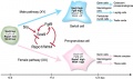
 Ovarian development model.jpg 1,200 × 1,036; 445 KB
 Ovarian developmental genes.jpg 600 × 490; 82 KB
 Ovarian follicle growth in vitro.jpg 1,000 × 769; 73 KB
 Ovarian follicular steroid synthesis.jpg 1,200 × 645; 89 KB
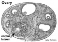
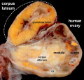
 Ovary corpus luteum.jpg 2,178 × 1,137; 376 KB
 Ovary follicle 01.jpg 450 × 600; 112 KB
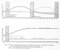
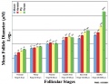 Ovary follicle size graph.jpg 1,057 × 820; 102 KB
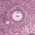
 Ovary Histology - tunica albuginea.jpg 1,280 × 1,024; 336 KB
 Ovary histology 001.jpg 1,280 × 1,024; 360 KB
 Ovary histology 002.jpg 1,280 × 1,024; 270 KB
 Ovary histology 003.jpg 1,280 × 1,024; 337 KB
 Ovary histology 004.jpg 1,280 × 1,024; 401 KB
 Ovary histology 005.jpg 1,280 × 1,024; 354 KB
 Ovary histology 006.jpg 1,280 × 1,024; 424 KB
 Ovary histology 007.jpg 1,280 × 1,024; 336 KB
 Ovary histology 008.jpg 1,280 × 1,024; 264 KB
 Ovary histology 061.jpg 1,280 × 1,024; 438 KB
 Ovary histology 061a.jpg 800 × 640; 200 KB
 Ovary histology 061c.jpg 400 × 320; 56 KB
 Ovary histology with chemotherapy.jpg 977 × 872; 232 KB
 Ovary oocyte size graph.jpg 1,057 × 820; 114 KB
 Ovary- atretic follicle 01.jpg 793 × 595; 225 KB
 Ovary- atretic follicle 02.jpg 600 × 450; 139 KB
 Ovary- atretic follicle 03.jpg 790 × 593; 202 KB
 Ovary- atretic follicle 04.jpg 600 × 450; 128 KB
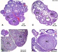
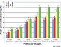
 Ovary- follicle stages.jpg 600 × 337; 45 KB
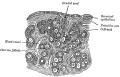
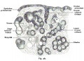
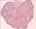
 Ovary- histology overview.jpg 861 × 646; 160 KB
 Ovary- histology secondary follicle 01.jpg 1,000 × 800; 293 KB
 Ovary-human-follicle.jpg 492 × 1,000; 103 KB
 Ovary10x.jpg 480 × 400; 53 KB
 Ovary5x.gif 480 × 400; 162 KB
 Pouchet1847 plate03.jpg 1,721 × 2,000; 456 KB
 Pouchet1847 plate04.jpg 1,787 × 2,000; 388 KB
 Pouchet1847 plate05.jpg 1,659 × 2,000; 334 KB
 Pouchet1847 plate06.jpg 1,616 × 2,000; 368 KB
 Rat ovary follicle development 01.jpg 1,200 × 1,035; 201 KB
 Rat ovary histology 01.jpg 1,200 × 938; 274 KB
 Simkins1928 plate01.jpg 1,574 × 2,003; 237 KB
 Simkins1928 plate02.jpg 1,464 × 2,126; 223 KB
 Simkins1928 plate03.jpg 1,551 × 2,086; 293 KB
 Simkins1928 plate04.jpg 1,543 × 2,026; 219 KB
 Simkins1928 plate05.jpg 1,281 × 2,111; 154 KB
 Simkins1928 plate06.jpg 1,587 × 2,088; 277 KB
 Simkins1928 plate07.jpg 1,540 × 2,096; 239 KB
 Simkins1928 plate08.jpg 1,558 × 1,797; 220 KB
 Simkins1928 plate09.jpg 1,548 × 2,096; 263 KB
 Simkins1928 plate10.jpg 1,565 × 1,386; 140 KB
 Ultrasound uterine and ovarian vascularity.jpg 531 × 780; 132 KB
 Week1 summary.jpg 1,000 × 747; 107 KB
 XXhpgaxis.gif 300 × 495; 22 KB
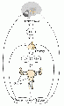
 XXhpgaxis.jpg 300 × 495; 24 KB


