Category:Notochord: Difference between revisions
mNo edit summary |
mNo edit summary |
||
| Line 1: | Line 1: | ||
This {{Embryology}} category shows media and pages related to [[Notochord|notochord]] development. | This {{Embryology}} category shows media and pages related to [[Notochord|notochord]] development. | ||
:'''Links:''' [[Notochord|Notochord]] | [[Paper - The Later Development of the Notochord in Mammals]] | :'''Links:''' [[Notochord|Notochord]] | [[Paper - The Later Development of the Notochord in Mammals|1908 Notochord]] | ||
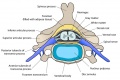
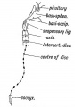
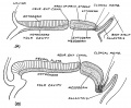
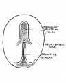
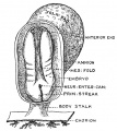
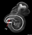
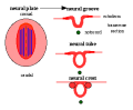
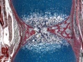
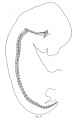
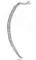
The notochord (axial mesoderm) is a rod of cells lying in the midline of the trilaminar embryo mesoderm layer ventral to the neural tube. Thought to have at least 2 early roles in development and later roles in patterning surrounding tissues. 1. Mechanical, influencing the folding of the early embryo; 2. Morphogenic, secreting sonic hedgehog a protein which regulates the development of surrounding tissues (neural plate, somites, endoderm and other organs). In humans, the notochord forms in week 3 and is eventually lost during the formation of the vertebral column. | The notochord (axial mesoderm) is a rod of cells lying in the midline of the trilaminar embryo mesoderm layer ventral to the neural tube. Thought to have at least 2 early roles in development and later roles in patterning surrounding tissues. 1. Mechanical, influencing the folding of the early embryo; 2. Morphogenic, secreting sonic hedgehog a protein which regulates the development of surrounding tissues (neural plate, somites, endoderm and other organs). In humans, the notochord forms in week 3 and is eventually lost during the formation of the vertebral column. | ||
Latest revision as of 13:06, 24 September 2015
This Embryology category shows media and pages related to notochord development.
- Links: Notochord | 1908 Notochord
The notochord (axial mesoderm) is a rod of cells lying in the midline of the trilaminar embryo mesoderm layer ventral to the neural tube. Thought to have at least 2 early roles in development and later roles in patterning surrounding tissues. 1. Mechanical, influencing the folding of the early embryo; 2. Morphogenic, secreting sonic hedgehog a protein which regulates the development of surrounding tissues (neural plate, somites, endoderm and other organs). In humans, the notochord forms in week 3 and is eventually lost during the formation of the vertebral column.
Pages in category 'Notochord'
The following 23 pages are in this category, out of 23 total.
P
R
Media in category 'Notochord'
The following 32 files are in this category, out of 32 total.
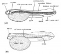
 Cervical vertebra.jpg 767 × 514; 71 KB
 Gray0065.jpg 600 × 345; 39 KB
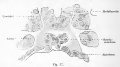
 Human notochord development theories 1.jpg 1,280 × 496; 64 KB
 Keith1902 fig015a.jpg 971 × 600; 74 KB
 Keith1902 fig117.jpg 561 × 800; 46 KB
 Keith1921 fig035.jpg 984 × 808; 143 KB
 Keith1921 fig036.jpg 547 × 686; 42 KB
 Keith1921 fig037.jpg 612 × 687; 102 KB
 Keith1921 fig040.jpg 901 × 806; 138 KB
 Kollmann057.jpg 845 × 469; 49 KB
 Mouse E11 Foxf1.jpg 1,420 × 1,143; 162 KB
 Mouse E11 Sox2 and Nkx2.1.jpg 2,229 × 720; 176 KB
 Mouse E9 Foxf1.jpg 1,420 × 1,143; 142 KB
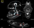
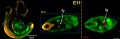
 Mouse embryo E11 HNF3beta notochord marker 01.jpg 2,245 × 829; 129 KB
 Mouse embryo E11 HNF3beta notochord marker 02.jpg 913 × 1,000; 68 KB
 Mouse embryo E11 HNF3beta notochord marker 03.jpg 913 × 1,000; 56 KB
 Mouse embryo E11 HNF3beta notochord marker 04.jpg 913 × 1,000; 55 KB
 Neuralplate cartoon.png 343 × 284; 5 KB
 Notochordal interaction with nucleus pulposus.jpg 478 × 360; 92 KB
 Ossification endochondral 01.jpg 817 × 613; 198 KB
 Ossification endochondral 1.jpg 750 × 1,000; 147 KB
 Ossification endochondral 1c.jpg 300 × 400; 32 KB
 Shh frog notochord 1.jpg 150 × 150; 8 KB
 Stage 13 image 056.jpg 1,000 × 516; 102 KB
 Stage 13 image 057.jpg 1,000 × 511; 99 KB
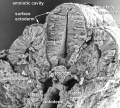 Stage11 sem100.jpg 1,000 × 898; 109 KB
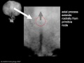 Stage7 axial process.jpg 500 × 375; 11 KB
 Williams1908-fig17.jpg 982 × 1,500; 145 KB
 Williams1908-fig18.jpg 921 × 1,500; 89 KB
 Williams1908-fig19.jpg 981 × 1,500; 104 KB
 Williams1908-fig20.jpg 920 × 1,500; 96 KB
 Xenopus FoxA4 model.jpg 1,192 × 565; 104 KB


