Category:Jan Florian

This category lists Embryology pages and media related to the historic Czech embryologist Jan Florian (1897 - 1942).
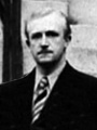
He held the chair of embryology at Masaryk University in Brno and published several key papers on early human development, along with J.P. Hill, in London. In 1942 he protested the Nazi decision to shut down Czech universities and was executed in Mauthausen.
External Links
External Links Notice - The dynamic nature of the internet may mean that some of these listed links may no longer function. If the link no longer works search the web with the link text or name. Links to any external commercial sites are provided for information purposes only and should never be considered an endorsement. UNSW Embryology is provided as an educational resource with no clinical information or commercial affiliation.
- Masaryk University - A Brief History of the Faculty of Medicine
Subcategories
This category has the following 3 subcategories, out of 3 total.
Pages in category 'Jan Florian'
The following 12 pages are in this category, out of 12 total.
F
P
- Paper - A Young Human Embryo (Embryo Dobbin) with Head-Process and Prochordal Plate
- Paper - An early human embryo (no. 1285, Manchester Collection) with capsular attachment of the connecting stalk (1935)
- Paper - An Early Human Embryo (No. 1285, Manchester Collection), with Capsular Attachment of the Connecting Stalk
- Paper - Further note on the pro-chordal plate in man
- Paper - The Early Development of Man, with Special Reference to the Development of the Mesoderm and Cloacal Membrane
- Paper - The Formation of the Connecting Stalk and the Extension of the Amniotic Cavity towards the Tissue of the Connecting Stalk in Young Human Embryos
Media in category 'Jan Florian'
The following 41 files are in this category, out of 41 total.
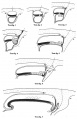 Florian1930-text-fig01-07.jpg 1,200 × 1,862; 261 KB
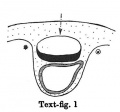 Florian1930-text-fig01.jpg 384 × 359; 19 KB
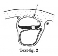 Florian1930-text-fig02.jpg 394 × 377; 20 KB
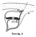 Florian1930-text-fig03.jpg 407 × 425; 25 KB
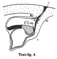 Florian1930-text-fig04.jpg 423 × 441; 31 KB
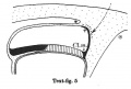 Florian1930-text-fig05.jpg 659 × 444; 40 KB
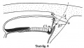 Florian1930-text-fig06.jpg 791 × 445; 50 KB
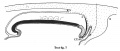 Florian1930-text-fig07.jpg 1,200 × 512; 78 KB
 Florian1930-text-fig08-09.jpg 1,000 × 1,347; 165 KB
 Florian1930-text-fig08.jpg 777 × 454; 49 KB
 Florian1930-text-fig09.jpg 1,000 × 903; 116 KB
 Florian1930-text-fig10.jpg 1,000 × 801; 68 KB
 Florian1930-text-fig11.jpg 1,739 × 1,000; 174 KB
 Florian1933 fig01.jpg 532 × 605; 38 KB
 Florian1933 fig02.jpg 580 × 569; 37 KB
 Florian1933 fig03.jpg 602 × 593; 40 KB
 Florian1933 fig04.jpg 754 × 610; 50 KB
 Florian1933 fig05.jpg 682 × 605; 51 KB
 Florian1933 fig06.jpg 731 × 584; 55 KB
 Florian1933 fig07.jpg 1,059 × 590; 60 KB
 Florian1933 fig08.jpg 1,109 × 520; 55 KB
 Florian1935 chorionic vesicle fig11.jpg 1,009 × 952; 248 KB
 Florian1935 fig01.jpg 774 × 405; 115 KB
 Florian1935 fig02.jpg 790 × 405; 117 KB
 Florian1935 fig03.jpg 800 × 455; 130 KB
 Florian1935 fig04.jpg 798 × 443; 120 KB
 Florian1935 fig05.jpg 803 × 227; 58 KB
 Florian1935 fig06.jpg 770 × 573; 145 KB
 Florian1935 fig07.jpg 597 × 812; 206 KB
 Florian1935 fig08.jpg 691 × 480; 114 KB
 Florian1935 fig09.jpg 510 × 765; 168 KB
 Florian1935 fig10.jpg 464 × 721; 157 KB
 Florian1935 fig11.jpg 1,280 × 1,427; 443 KB
 Florian1935 plate01.jpg 1,481 × 1,825; 529 KB
 Florian1935 plate02.jpg 1,481 × 1,806; 578 KB
 Florian1935 plate03.jpg 1,481 × 2,000; 735 KB
 Florian1935 textfig01.jpg 1,120 × 1,202; 136 KB
 Florian1935 textfig02.jpg 1,579 × 2,000; 342 KB
 Florian1935 textfig03.jpg 1,579 × 2,000; 212 KB
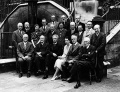 International Institute of Embryology, London 1938.jpg 1,000 × 763; 224 KB
 Jan Florian.jpg 300 × 400; 14 KB


