Brain Awareness Week 2012
From Embryology
Welcome to Brain Development
| width=320px|height=260px|controller=false|autoplay=true</qt> | In today's demonstration we will be looking at how the brain develops from a simple tube into the complex folded structure you will be seeing (and using) today.
|
Here is Human Development

Here is how the human nervous system grows
|
|||||
| Week 3 | Week 4 to 5 | Week 5 | Week 8 | Week 13 to 21 | Adult Human |
| Neural Plate | Neural Tube | Simple Tube | Central Nervous | Fetal Brain | Brain Slices |
Here is a developing mouse nervous system
| width=336px|height=415px|controller=true|autoplay=false</qt> |
This movie shows a mouse 11.5 days old. (Mouse development takes 21 days)
Red - brain
|
|
It begins as a Plate
| width=316px|height=520px|controller=true|autoplay=false</qt> |
|

|
That folds to a Tube
| width=480px|height=500px|controller=true|autoplay=false</qt> |
The human embryo is now 4 weeks old and sits on top of a big yolk sac.
|

|
The tube then Closes at each End

|

|
Why are these important? |


The brain end of the tube forms 3 Vesicles
The tube expands at the brain end to form three vesicle (sac or bubble) regions.
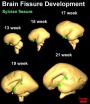
Fetal brain growth

This shows the growth of the brain and the fluid-filled space within the brain (the red bar is 1 cm).
- The brain goes from having a smooth surface to begin to fold or "wrinkle".
- The fluid space is filled with cerebral-spinal fluid or CSF.