Brain Awareness Week 2012: Difference between revisions
| Line 26: | Line 26: | ||
|- | |- | ||
| Week 3 | | Week 3 | ||
| Week 4 | | Week 4 to 5 | ||
| Week 5 | | Week 5 | ||
| Week 8 | | Week 8 | ||
| Week 13 | | Week 13 to 21 | ||
| Adult Human | | Adult Human | ||
|- | |- | ||
Revision as of 18:32, 8 March 2012
Welcome to "Brain Development"
| width=320px|height=260px|controller=false|autoplay=true</qt> | In today's demonstration we will be looking at how the brain develops from a simple tube into the complex folded structure you will be seeing (and using) today.
|
Here is Human Development

Here is how the human nervous system grows
|
|||||
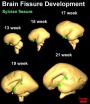
| Week 3 | Week 4 to 5 | Week 5 | Week 8 | Week 13 to 21 | Adult Human |
| Neural Plate | Neural Tube | Simple Tube | Central Nervous | Fetal Brain | Brain Slices |
Here is a developing mouse nervous system
| width=336px|height=415px|controller=true|autoplay=false</qt> |
This movie shows a mouse 11.5 days old. (Mouse development takes 21 days)
Red - brain
|
|
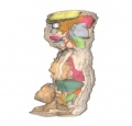
| <Flowplayer height="588" width="560" autoplay="true">Stage13_3dCNSlarge.flv</Flowplayer> | 3 Dimensional Reconstruction
Based upon a serial reconstruction from individual embryo slice images. (6 mm pig embryo, approximately Human day 32, Carnegie Stage 13/14 embryo) The ectoderm contributes the neural tube structures of the central nervous system. Below the animation is a more complete description of this system.
|
| <Flowplayer height="415" width="400" autoplay="true">Stage22 CNS.flv</Flowplayer> | 3 Dimensional Reconstruction
Based upon a serial reconstruction from individual embryo slice images (27 mm Human embryo, Carnegie Stage 22 approximate day 56).
Colour code:
These 3d movies were part of the UNSW Medical degree Independent Learning Project (ILP) prepared by Aashish Kumar (2006).
|