Book - The Elements of Embryology - Figures
| Embryology - 26 Apr 2024 |
|---|
| Google Translate - select your language from the list shown below (this will open a new external page) |
|
العربية | català | 中文 | 中國傳統的 | français | Deutsche | עִברִית | हिंदी | bahasa Indonesia | italiano | 日本語 | 한국어 | မြန်မာ | Pilipino | Polskie | português | ਪੰਜਾਬੀ ਦੇ | Română | русский | Español | Swahili | Svensk | ไทย | Türkçe | اردو | ייִדיש | Tiếng Việt These external translations are automated and may not be accurate. (More? About Translations) |
Foster M. Balfour FM. Sedgwick A. and Heape W. The Elements of Embryology (1883) Vol. 1. (2nd ed.). London: Macmillan and Co.
| Historic Disclaimer - information about historic embryology pages |
|---|
 |
Volume 1 - The History of the Chick

- The structure of the hen's egg, and the changes which take place up to the beginning of incubation
- A brief summary of the whole history of incubation
- The changes which take place during the first day of incubation
- The changes which take place during the first half of the second day
- The changes which take place during the second half of the second day
- The changes which take place during the third day
- The changes which take place during the fourth day
- The changes which take place on the fifth day
- From the sixth day to the end of incubation
- Appendix
Volume 2 - The History of the Mammalian Embryo
- General Development of the Embryo
- Embryonic Membranes and Yolk-Sac
- Organs from Epiblast
- Organs from Mesoblast
- Alimentary Canal
- Appendix
Volume 1 - Figures
The History of the Chick
The structure of the hen's egg, and the changes which take place up to the beginning of incubation
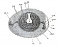
Fig. 1 Diagrammatic section of an unincubated fowl's egg (modified from allen thomson).
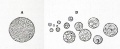
Fig. 2 Yellow yolk-sphere filled with fine granules.
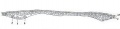
Fig. 3. Section of a blastoderm of a fowl's egg at the commencement of incubation.

Fig. 4. Section through the germinal disc of the ripe ovarian ovum of a fowl while yet enclosed in its capsule.
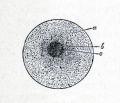
Fig. 5. Diagram of the ovum. (from Gegenbaur.)

Fig. 6. surface views of the early stages of the segmentation in a fowl's egg. (a and c after Coste.)
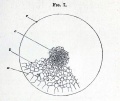
Fig. 7. Surface view of the germinal disc of a hen's egg during the later stages of segmentation.
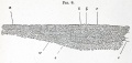
Fig. 8. Section of the germinal disc of a fowl during the later stages of segmentation.


A brief summary of the whole history of incubation
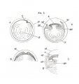
Fig. 9, A to D series introduces embryo body, yolk-sac, amnion and allantois.

Fig. 9, E to K series introduces embryo body, yolk-sac, amnion and allantois.

Fig. 9, L to N series introduces embryo body, yolk-sac, amnion and allantois. allantois.
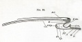
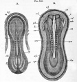
Fig. 10. Longitudinal section through the axis of an embryo.
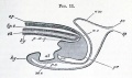
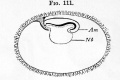
Fig. 11. Longitudinal section posterior end at time of allantois formation.


The changes which take place during the first day of incubation
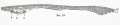
Fig. 12. Section of a blastoderm of a fowl's egg at the commencement of incubation.

Fig. 13. Transverse section through the blastoderm of a chick before the appearance of the primitive streak.
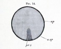
Fig. 14. Area pellucida of a very young blastoderm of a chick.

Fig. 15. Transverse section through a blastoderm.
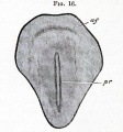
Fig. 16. Surface view of the area pellucida after the formation of the primitive groove.
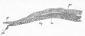
Fig. 17. Transverse section through the front end op the primitive streak of a blastoderm.

Fig. 18. Longitudinal section through the axial line of the primitive streak.

Fig. 19. Transverse section prior to formation of medullary groove and notochord.

Fig. 20. Transverse section at the time of notochord formation, before appearance of medullary groove.

Fig. 21. Transverse section of a blastoderm incubated for 18 hours.
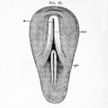
Fig. 22. View of the pellucid area of a blastoderm of 18 hours.

Fig. 23. Dorsal view of the hardened area pellucida of a chick with five mesoblastic somites.
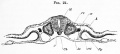
Fig. 24. Transverse section through the dorsal region op an embryo of the second day.
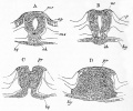
Fig. 25. Transverse sections neurenteric passage in duck embryo twenty-six mesoblastic somites.

Fig. 26. Diagrammatic longitudinal section through the posterior end of an embryo bird at the time of the formation of the allantois.


The changes which take place during the first half of the second day
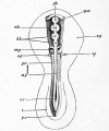
Fig. 27. Embryo of the chick between thirty and thirty-six hours, viewed from above as an opaque object.
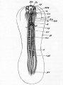
Fig. 28. an embryo chick of about thirty-six hours, viewed from below as a transparent object.
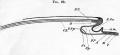
Fig. 29. Diagrammatic longitudinal section through the axis of an embryo.
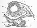
Fig. 30. Transverse section through the posterior part of the head of an embryo chick of thirty hours.
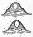
Fig. 31. Two sections of a thirty-six hours' embryo heart shortly after its formation. a is the anterior section.
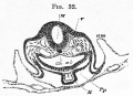
Fig. 32. Transverse section at the end of the second day through the bulbus arteriosus. (Copied from His.)
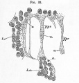
Fig. 33. Surface view from below of the posterior end pellucid area of a thirty-six hours' chick.


The changes which take place during the second half of the second day
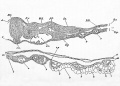
Fig. 34. Transverse section through the dorsal region of an embryo of 45 hours.
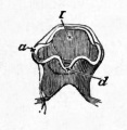
Fig. 35. Head of a chick at the end of the second day viewed from below as a transparent object. (Copied from Huxley).


The changes which take place during the third day
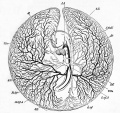
Fig. 36. Circulation of the yolk-sack at the end of the third day of incubation.
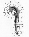
Fig. 37. chick of the third day (fifty-four hours) viewed from underneath as a transparent object.
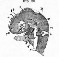
Fig. 38. Head of a chick of the third day viewed sideways as a transparent object. (From Huxley.)
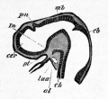
Fig. 39. Longitudinal section through the brain of a young pristiurus embryo.
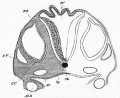
Fig. 40. Section through the hind-brain of a chick at the end of the third day of incubation.
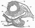
Fig. 41. Transverse section through the posterior part of the head of an embryo chick of thirty hours.
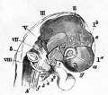
Fig. 42. Head of an embryo chick of the third day (seventy five hours) viewed sideways as a transparent object. (From Huxley.)
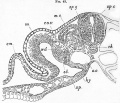
Fig. 43. Transverse section through the trunk of a duck embryo with about twenty-four mesoblastic somites.

Fig. 44. Transverse section through the trunk of a young embryo of a dog-flsh.
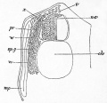
Fig. 45. Section through the dorsal region of an embryo dog-fish.
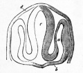
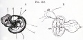
Fig. 46. Section through the head of an embryo teleostean, to shew the formation of the optic vesicles.
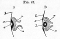
Fig. 47. Sections illustrating the formation of the eye. (After Kemak.)
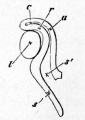
Fig. 48. Section of the eye and the optic nerve at an early stage (from Lieberkuhn).

Fig. 49. The eye of the chick of about the third day as seen when the head is viewed from underneath as a transparent object.
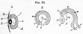
Fig. 50. Section of the eye and the optic nerve at an early stage.


Volume 2 - Figures
The History of the Mammalian Embryo
General Development of the Embryo
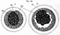
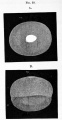
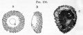
Fig. 95. Optical sections of a rabbit's ovum at two stages closely following upon the segmentation.

Fig. 96. Rabbit's ovum between 70 90 hours after impregnation.
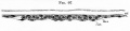
Fig. 97. Section through the nearly circular embryonic area of a rabbit ovum of six days.

Fig. 98. Section through the blastoderm of a rabbitt on the seventh day: taken in front of the primitive streak


Embryonic Membranes and Yolk-Sac
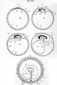
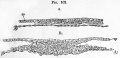
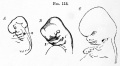
Fig. 114. Five diagrammatic figures illustrating the formation of the foetal membranes of a mammal. (From Kolliker.)
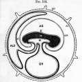
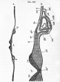
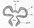
Fig. 115. Diagram of the foetal membranes op a mammal. (From Turner.)

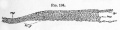
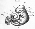
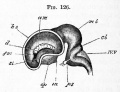
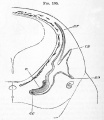
Fig. 116. Diagrammatic longitudinal section of a rabbit's ovum at an advanced stage of pregnancy. (From Kolliker after Bischoff.)
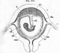
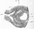
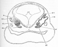
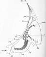
Fig. 117. Diagrammatic section of pregnant human uterus with contained foetus. (From Huxley after Longet.)
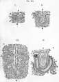
Fig. 118. Histology of the placenta. diagrammatic representations of the minute structure of the placenta. (From Turner.)
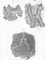
Fig. 118. Histology of the placenta. diagrammatic representations of the minute structure of the placenta. (From Turner.)


Organs from Epiblast

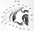
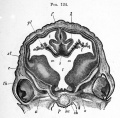
Fig. 119. Longitudinal section through the brain of a chick of ten days
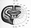


Organs from Mesoblast

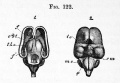
Fig. 134. Longitudinal section through the vertebral column of an eight weeks' human embryo in the thoracic region.
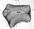
Fig. 135. Longitudinal section through the intervertebral ligament and adjacent parts of two vertebra from the thoracic region of an advanced embryo of a sheep.
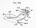
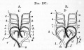
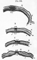
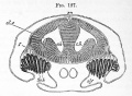
Fig. 137. Diagrams illustrating the metamorphosis of the arterial arches in a bird A. and a mammal B.
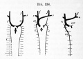
Fig. 138. Diagram of the development of the paired venous system of mammals (man).

Fig. 139. Diagram of the chief venous trunks of man.
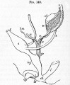
Fig. 140. Diagram of the urinogenital organs of a mammal at an early stage.


Alimentary Canal
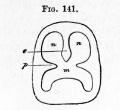
Fig. 141. Diagram shewing the division of the primitive buccal cavity into the respiratory section above and the true mouth below

Glossary Links
- Glossary: A | B | C | D | E | F | G | H | I | J | K | L | M | N | O | P | Q | R | S | T | U | V | W | X | Y | Z | Numbers | Symbols | Term Link
Cite this page: Hill, M.A. (2024, April 26) Embryology Book - The Elements of Embryology - Figures. Retrieved from https://embryology.med.unsw.edu.au/embryology/index.php/Book_-_The_Elements_of_Embryology_-_Figures
- © Dr Mark Hill 2024, UNSW Embryology ISBN: 978 0 7334 2609 4 - UNSW CRICOS Provider Code No. 00098G