Book - Stoehr's Histology (1906): Difference between revisions
mNo edit summary |
mNo edit summary |
||
| Line 1: | Line 1: | ||
{{Lewis1906 header}} | {{Lewis1906 header}} | ||
{| class="wikitable mw-collapsible mw-collapsed" | |||
See also the second edition | ! Online Editor | ||
|- | |||
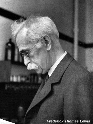
| [[File:Mark_Hill.jpg|90px|left]] This historic 1906 textbook translated by [[Embryology History - Frederic Lewis|Frederic T. Lewis]] from the original German of Stöhr, describes histology organised upon an embryological basis. Note that linked terms within the textbook go to the modern pages and the "Online Editor" sections provide additional information.<br> | |||
See also the 1913 second edition: {{Ref-LewisStöhr1913}} | |||
<br><br> | |||
[[Media:1906 A Text-book of Histology - Arranged Upon an Embryological Basis.pdf|PDF version]] | [https://archive.org/details/atextbookhistol00schugoog Internet Archive] | |||
<br> | |||
[[Historic Embryology Textbooks]] | |||
<br> | |||
|} | |||
=Stöhr's Histology= | =Stöhr's Histology= | ||
[[File:Stoehr's Histology 1906 titlepage.jpg|thumb|400px|Stoehr's Histology titlepage]] | [[File:Stoehr's Histology 1906 titlepage.jpg|thumb|400px|Stoehr's Histology titlepage]] | ||
| Line 13: | Line 21: | ||
Assistant Professor of Embryology at the Harvard Medical School | Assistant Professor of Embryology at the Harvard Medical School | ||
From the Twelfth German Edition | From the Twelfth German Edition | ||
By | By | ||
| Line 23: | Line 29: | ||
Professor of Anatomy at the University Of Würzburg | Professor of Anatomy at the University Of Würzburg | ||
Sixth American Edition | Sixth American Edition | ||
With 450 illustrations | With 450 illustrations | ||
Philadelphia | Philadelphia | ||
P. Blakiston's Son & Co. | P. Blakiston's Son & Co. 1012 Walnut Street 1906 | ||
1012 Walnut Street 1906 | |||
Copyright, 1903, by Dr. Alfred Schaper | Copyright, 1903, by Dr. Alfred Schaper | ||
Copyright, 1906, by Estatr of Dr. Alfred Schaper | Copyright, 1906, by Estatr of Dr. Alfred Schaper | ||
Press of WM. Fell Company, 1820-24 Sandom Street, PHILADELPHIA, PA | Press of WM. Fell Company, 1820-24 Sandom Street, PHILADELPHIA, PA | ||
==Note== | ==Note== | ||
In the new edition of the American translation of my hand-book a number of additions and changes have been made by the translator with my permission. It is therefore reasonable that I should not take the same responsibility for the translation as for the text of the German original, and I would ask those of my colleagues who wish to question the correctness of my assertions in their papers, to convince themselves, by making comparisons with my last German edition, that the paragraphs in question were written by me. | In the new edition of the American translation of my hand-book a number of additions and changes have been made by the translator with my permission. It is therefore reasonable that I should not take the same responsibility for the translation as for the text of the German original, and I would ask those of my colleagues who wish to question the correctness of my assertions in their papers, to convince themselves, by making comparisons with my last German edition, that the paragraphs in question were written by me. | ||
Revision as of 10:37, 9 April 2020
| Embryology - 26 Apr 2024 |
|---|
| Google Translate - select your language from the list shown below (this will open a new external page) |
|
العربية | català | 中文 | 中國傳統的 | français | Deutsche | עִברִית | हिंदी | bahasa Indonesia | italiano | 日本語 | 한국어 | မြန်မာ | Pilipino | Polskie | português | ਪੰਜਾਬੀ ਦੇ | Română | русский | Español | Swahili | Svensk | ไทย | Türkçe | اردو | ייִדיש | Tiếng Việt These external translations are automated and may not be accurate. (More? About Translations) |
Lewis FT. Stoehr's Histology. (1906) P. Blakiston's Son & Co., Philadelphia.
| Stoehr's Histology 1906: 1 Microscopic Anatomy | 1-1 Cytology | 1-2 General Histology | 1-3 Special Histology | 2 Preparation of Specimens | Figures | Histology | Embryology History |
| Historic Disclaimer - information about historic embryology pages |
|---|
 |
- Note - This historic textbook is currently in an early stage of online preparation. (this notice removed when completed)
| Online Editor |
|---|
See also the 1913 second edition: Lewis FT. and Stöhr P. A Text-book of Histology Arranged upon an Embryological Basis. (1913) P. Blakiston’s Son and Co., 539 pp., 495 figs.
|
Stöhr's Histology

Arranged Upon An Embryological Basis
By
Assistant Professor of Embryology at the Harvard Medical School
From the Twelfth German Edition
By
Dr. Philipp Stoehr (Stöhr)
Professor of Anatomy at the University Of Würzburg
Sixth American Edition
With 450 illustrations
Philadelphia
P. Blakiston's Son & Co. 1012 Walnut Street 1906 Copyright, 1903, by Dr. Alfred Schaper Copyright, 1906, by Estatr of Dr. Alfred Schaper Press of WM. Fell Company, 1820-24 Sandom Street, PHILADELPHIA, PA
Note
In the new edition of the American translation of my hand-book a number of additions and changes have been made by the translator with my permission. It is therefore reasonable that I should not take the same responsibility for the translation as for the text of the German original, and I would ask those of my colleagues who wish to question the correctness of my assertions in their papers, to convince themselves, by making comparisons with my last German edition, that the paragraphs in question were written by me.
Philipp Stohr.
Preface
The need of a text-book of histology arranged upon an embryological basis has long been felt. At the Harvard Medical School this need has been urgent. There Professor Schaper, the editor of the five previous American editions of Stohr's Histology planned such a book, and after his return to Germany its preparation was begun. It is greatly to be regretted that at the time of his death the work was only commenced, for there was promise of a notable production.
When the writer was informed that Professor Stohr had given generous permission to adapt a new edition of his Histology to American needs it was decided to rearrange the book upon an embryological plan. This has been accomplished with the loss of some characteristic features of the German edition, for which the added material will, it is hoped, make compensation. Thus in order to have space for describing the controlling developmental features of the organs, and for presenting their adult structure somewhat more fully, the directions for preparing sections have been reduced to the minimum. These may be supplemented by directions in the class room; and for the small proportion of students who intend to practice elaborate microscopical methods, a special text-book may be recommended. It is not essential that a physician should be familiar with the details of many staining processes, but the structure of the adult organs and the developmental possibilities of their constituent tissues must be known.
The nomenclature adopted is that published by the committee of the German Association of Anatomists m 1895 (-^rcA. /. Anat, u. Phys.; Anat, Abth.; Supplement-Band) J and which is now widely used. It is founded upon the sound principle that the name of a structure should be the simplest possible descriptive Latin term or phrase. Since the Latin names may be translated into the various modem languages the nomenclature is international. Moreover a large number of the names are conmionly used in their Latin forms. Personal names have been discarded (except Wolffian and Milllertan), thus greatly assisting the student. It is obviously easier to learn intestinal glands , duodenal glands, parotid duct, etc., rather than Lieberkiihn's glands, Brunner's glands, Stenson's duct, and the like. It has been estimated that five thousand synonyms have been rejected and are to be removed from the anatomist's vocabulary as soon as possible. In the following pages the more common of the rejected names have been placed in square brackets, [ ]. However difficult it may be for the older anatomists to conform to this nomenclature, it seems clearly a duty to the overworked medical students to adopt it.
Excellent as the German nomenclature is, as a whole, it is not beyond improvement, and it may be desirable for a conMnittee of the Association of American Anatomists to publish in their English forms a corresponding list of names.[1] As few changes as possible should be made, but it is certain , for example, that the ventral surface of the body will not be called anterior , or the dorsal surface posterior. In the following pages anterior always means toward the head. Conunon general terms should be made even more specific. For instance, it is questionable whether follicle (Latin, a small leather bag, a husk or shell) should be applied to anything other than closed cysts like the follicles of the ovary and thyreoid gland. Its application by the Germans to the sheath of the hair and by many Americans to solid nodules of lymphoid tissue may lead the student to wonder if ** follicle" is not a colloquial rather than a scientific term.
The attention of all students should be called to the American Journal of Anatomy, the quarterly publication of the Association of American Anatomists, which contains the results of current American anatomical and histological investigations. It probably aflfords the most satisfactory means by which a physician may keep in touch with these sciences.
The writer has many acknowledgments to make for help received. Messrs. P. Blakiston's Son & Co., and Mr. William T. Oliver, the artist who has drawn the more elaborate of the new figures, have rendered all the assistance possible. Members of several departments at the Harvard Medical School have given valuable advice, and Dr. G. H. Wright, Assistant in Dental Histology, has arranged a considerable portion of the section on the teeth. It is a privilege to present for the first time in a textbook, the discoveries of Dr. James H. Wright regarding the origin of blood plates. His remarkable conclusion that they are fragments of pseudopodia of the giant cells seems established beyond doubt by an examination of his specimens.
Finally it is a pleasure to record that after studying histology and embryology under Professor Charles S. Minot, the writer has for several years enjoyed the closest association with him in his scientific work. The results of such unusual privilege should be found reflected in this edition of Professor Stohr's Histology.
Frederic T. Lewis.
Cambridge, Massachusetts,
September, 1906.
- ↑ The writer has since been informed that Messrs. Blakiston's Son & Co. have in press such a list prepared by Professor Barker and entitled "Anatomical Terminology" The orderly arrangement of these descriptive names makes the Latin list - and undoubtedly their English version also - an excellent means by which students may review anatomy.
| Other Texts by Frederic T. Lewis |
|---|
|
Contents
Part I. Microscopic Anatomy
I. Cytology
- The Cell,
- Protoplasm.
- Nucleus.
- Centrosome.
- Cell Wall.
- Form and Size OF Cells
- Vital Phenomena
- Amoeboid Motion.
- Formation and Reproduction of Cells
- Mitosis
- Amitosis
- Cytomorphosis
II. General Histology
- Histogenesis
- Segmentation and the Formation of the Germ Layers.
- The Fundamental Tissues.
- Epithelia
- Origin
- Shapes of Epithelial Cells.
- Number of Layers.
- Differentiation.
- Processes of Secretion.
- The Nature and Classification of Glands
- Mesenchymal Tissues
- Reticular Tissue.
- Mucous Tissue.
- Connective Tissue.
- Tendon
- Cartilage.
- Bone.
- Joints.
- Teeth (including the Ectodermal Enamel Organs).
- Muscle Tissue
- Smooth Muscle
- Cardiac Muscle
- Striated Muscle
- Nerve Tissue,
- Development of,
- The central tract.
- The spinal ganglia.
- The ventral roots.
- The sympathetic system.
- The cerebral nerves.
- Structure of Nerve fibers and nerves
- Structure of Sensory endings.
- Structure of Motor endings.
- Structure of Gangiia
- The spinal cord.
- Vascular Tissue
- Blood Vessels.
- Development
- Capillaries.
- Arteries.
- Veins.
- The heart.
- Lymphatic Vessels.
- Red corpuscles.
- White corpuscles.
- Blood plates.
- Plasma.
- Lymph.
III. Special Histology
- Blood Forming and Blood Destroying Organs
- Bone Marrow
- Lymph Nodules and Lymph Glands
- Haemolyroph Glands
- Spleen
- The Entodermal Tract
- The Mouth and Pharynx
- Development.
- Palatine tonsils.
- Thymus.
- Thyreoid gland.
- Parathyreoid glands.
- Glomus caroticum.
- Tongue.
- Oral and pharyngeal cavities.
- Glands of the oral cavity.
- The Digestive Tube
- Development.
- Oesophagus.
- Stomach.
- Small Intestine.
- Large Intestine.
- Rectum and Anus.
- The Liver
- The Pancreas
- The Respiratory Tract
- Development.
- Larynx.
- Trachea, Bronchi.
- Lungs.
- Urinary Organs
- Wolffian Body.
- Pronephros.
- Kidney.
- Renal pelvis and ureter.
- Bladder.
- Urethra (in the female)
- Male Genital Organs
- Development.
- Testis.
- Epididymis.
- Ductus deferens.
- Seminal Vesicles and Ejaculatory Ducts.
- Appendices and Paradidymis.
- Prostate.
- Urethra and Penis.
- Female Genital Organs
- Development.
- Ovary.
- Epoophoron.
- Uterine Tubes.
- Uterus.
- Menstruation.
- Development of the decidual membranes.
- Structure of the membranes and placenta.
- Umbilical Cord.
- Vagina and External Genital Organs.
- Skin
- Nails.
- Hair.
- Sebaceous glands.
- Sweat glands.
- Mammary glands.
Suprarenal Glands
Brain and Sense Organs
Brain
Development.
Medulla oblongata.
Pons.
Cerebellum.
Hemispheres.
Hypophysis.
Pineal body.
Meninges.
- Eye
- Development.
- Retina.
- Optic nerve.
- Lens.
- Vitreous body.
- Tunica vasculosa.
- Tunica fibrosa.
- Vessels, chambers, and nerves.
- Eyelids.
- Lachrymal glands.
- Ear
- Development.
- Internal ear
- Sacculus.
- Utriculus.
- Semicircular ducts, and Cochlea.
- Middle ear.
- External ear.
- Nose
- Respiratory region.
- Olfactory region.
Part Ii. The Preparation and Examination of Microscopical Specimens
Part II. The Preparation and Examination of Microscopical Specimens
- Fresh Tissues
- Staining and Mounting
- Isolation.
- General Stains.
- Sectioning Fresh Material.
- Special Stains.
- Fixation.
- The Microscope.
- Decalcification.
- Drawings.
- Imbedding.
- Reconstructions.
| Historic Disclaimer - information about historic embryology pages |
|---|
|
| Stoehr's Histology 1906: 1 Microscopic Anatomy | 1-1 Cytology | 1-2 General Histology | 1-3 Special Histology | 2 Preparation of Specimens | Figures | Histology | Embryology History |
Cite this page: Hill, M.A. (2024, April 26) Embryology Book - Stoehr's Histology (1906). Retrieved from https://embryology.med.unsw.edu.au/embryology/index.php/Book_-_Stoehr%27s_Histology_(1906)
- © Dr Mark Hill 2024, UNSW Embryology ISBN: 978 0 7334 2609 4 - UNSW CRICOS Provider Code No. 00098G