Book - Russian Embryology (1750 - 1850) 5
| Embryology - 26 Apr 2024 |
|---|
| Google Translate - select your language from the list shown below (this will open a new external page) |
|
العربية | català | 中文 | 中國傳統的 | français | Deutsche | עִברִית | हिंदी | bahasa Indonesia | italiano | 日本語 | 한국어 | မြန်မာ | Pilipino | Polskie | português | ਪੰਜਾਬੀ ਦੇ | Română | русский | Español | Swahili | Svensk | ไทย | Türkçe | اردو | ייִדיש | Tiếng Việt These external translations are automated and may not be accurate. (More? About Translations) |
Blyakher L. History of embryology in Russia from the middle of the eighteenth to the middle of the nineteenth century (istoryia embriologii v Rossii s serediny XVIII do serediny XIX veka) (1955) Academy of Sciences USSR. Institute of the History of Science and Technology. Translation Smithsonian Institution (1982).
| Online Editor |
|---|
| This historic textbook by Bliakher translated from Russian, describes historic embryology in Russia between 1750 - 1850.
Publishing House of the Academy of Science USSR Moscow 1955 Translated from Russian Translated and Edited by: Dr. Hosni Ibrahim Youssef # Faculty of Veterinary Medicine Cairo University Dr. Boulos Abdel Malek Head of Veterinary Research Division NAMRU-3, Cairo Arab Republic of Egypt Published for The Smithsonian Institution and the National Science Foundation, Washington, D.C, by The Al Ahram Center for Scientific Translations 1982
The Smithsonian Institution and the National Science Foundation, Washington, D.C by The Al Ahram Center for Scientific Translations (1982)
|
| Historic Disclaimer - information about historic embryology pages |
|---|
| Pages where the terms "Historic" (textbooks, papers, people, recommendations) appear on this site, and sections within pages where this disclaimer appears, indicate that the content and scientific understanding are specific to the time of publication. This means that while some scientific descriptions are still accurate, the terminology and interpretation of the developmental mechanisms reflect the understanding at the time of original publication and those of the preceding periods, these terms, interpretations and recommendations may not reflect our current scientific understanding. (More? Embryology History | Historic Embryology Papers) |
Chapter 5 Wolff's Treatise "On the Formation of the Intestine"
This work, as already stated, represents one of the most important stages in the history of embryology. It was published and distributed in a limited edition in "New Commentaries of the Petersburg Academy of Science." It was known to few contemporaries, until Meckel translated it into German in 1812. After that, the biologists interested in the history of science acquainted themselves with Wolff's works. They turned to the distributed edition of the German translation of THEORIA GENERATION!. S, and these readers were able to form their own opinions about Wolff, concluding that his ideas were not always expressed in comprehensible language, were abstract, and were not very convincing discussions about development.
Wolff's work dedicated to the development of the intestine produced another impression completely. It is a systematic statement of thorough and impartial observations on the development of the chick embryo during the first four days of incubation. The conclusions which the investigator reached clearly arose from his informed facts and therefore were completely convincing. This truly classical work fairly deserved the high evaluation of the leader of embryological science, K. M. Baer.
The complete title of Wolff's work is "Observations carried out on incubated eggs, mainly on the formation of the intestine, and also on the false amnion and other parts of chick embryo that are still not observed today."! The work begins with a comparison of development in plants and animals. Wolff first noticed that the different parts from which the plant organisms are composed are extremely similar to each other, and therefore the ways of their development are easily established. Truly, as Wolff said, great depth of thought is not required to notice, especially in some plants, that in many composites in which the leaves gradually become smaller and more incomplete and adjoin nearer to each other the higher they are situated on the stem, at the end the last leaves, which are present directly under the flowers, turn out to be extremely small and are firmly adjacent to each other. They form the leaves of the calyx, and together constitute the calyx itself. Concerning the petals of the flower, Wolff did not doubt that they are also modified true leaves. He made the same conclusion for other parts, which in some flowers are transformed into petals, and vice versa. As a result of these observations Wolff concluded the following: if all parts of the plant, except the stem, are similar in their morphology to the leaves and are nothing other than modification of the latter, then it is not so difficult to formulate the theory of development in plants.
1. C. F. Wolff, DE FORMATIONE INTESTINORUM praecipue turn et de amnio spurio, aliisque partibus embryonis gallinacei, nondum visis observationes , in ovis incubatis institutae . NOVI COMMENT. ACAD. SCIENT. IMP. PETROPOLITANAE , v. XII (pro Anno 1766 - 1767), 1768, pp. 403 - 507, v. XIII (pro Anno 1768), 1769, pp. 478 - 530.
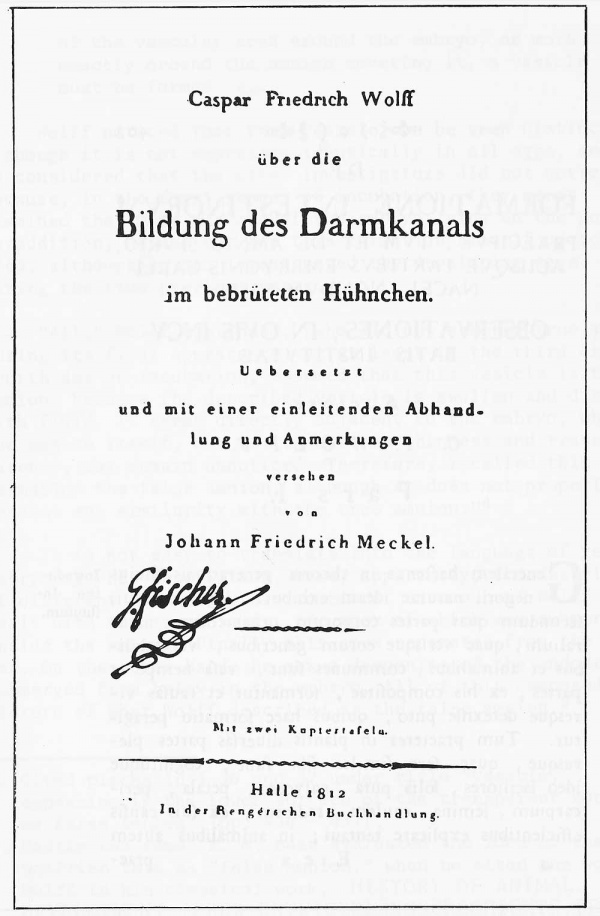
This title is situated in the first part of work, without an independent subtitle? second and third parts are called accordingly "Second part, Cardiac fossa; commissure of false amnion, lower fossa; beginning, increase and disappearance of these formations" and "Third part. Internal phenomena and false amnion, which at the same time stated the formation of mesentery, thorax, abdomen, wings and legs" . Meckel in his translation gave the work a briefer general title ON THE FORMATION OF THE INTESTINAL CANAL IN THE DEVELOPING CHICKEN (KASPAR FRIEDRICH WOLFF UBER DIE BILDUNG DES DARMKANALS IM BEBRUTETEN HUHNCHEN . Ubersetzt und mit einer einleitenden Abhandlung und Ammerkungen versehen von Johann Friederich Meckel, Halle, 1812) .
Repeatedly, it was noticed that Wolff pioneered the idea of unity of the morphological structure of plants, brought together to a general form in the leaf, which was later worked out in detail by Goethe. This side of Wolff's scientific interests is indirectly related to the basic direction of his activity.
Passing to the morphology of animals, Wolff noticed that the analogy cannot be established between the individual parts which exist in plants, and the different organs which exist in the animal body. The organized body of the animal is a very complicated whole; it originates as a result of interaction of many interconnected dependent causes. One of the forms of development characteristic for animals is similar to the development of plants; it appears in the formation of the extremities. Wolff wrote the following:
The extremities, i.e. wings and legs of birds, first appear in the form of protuberances , and then elongate; at their ends new protuberances appear which are the rudiments of the fingers, and gradually they acquire the form and structure of the formed leg and wing. Something similar occurs in vegetation, as the leaves of plants are formed; this common mode of formation I stated in the theory of generation. 3
This process of development is realized when, at the beginning, the nutritional fluid collects in the already existing parts and causes a protuberance to appear, which is the first rudiment of a new part. This fluid is gradually transformed into a more compact substance. Simultaneously with this, vessels appear in it, through which new nutritional juices flow. By this means a new part is organized, i.e. acquires structure. Similarly new protuberances appear in different places, being the rudiments of new parts.
2. DE FORMATIONE, p. 410.
3. Ibid., pp. 411 - 412.
In animals, other types of generation and development of parts of the body also exist. Thus, a different type of development than that just described characterizes the intestine, the fundamental object of Wolff's observations in the discussed work. Already in the beginning of the work ($ 2), Wolff confirmed that he could understand the initial stages of the genesis of the intestinal canal only because he traced the entire process of development of the intestine from the moment of its formation to its final completion. Wolff expressed a hope that his theory about formation of the intestinal canal would be acceptable for experienced naturalists, as it is nearly wholly taken from observations.
In another place (5 81) Wolff described his method of studying the developing eggs, which he considered the only suitable method, in order to make the description of the phenomena completely understandable; thus: "I selected the following order in the description of the phenomena in the incubated egg. From the beginning I described the phenomena at that time when they attain the highest development.... then I followed them to the moment of appearance, described the changes which occur from their first appearance to that time when their essence can be determined. Finally I explained the other changes, which occur in the period up to the full completion of the process."
Passing to the statement of the basic results of his work, Wolff turned attention to the previously insufficiently studied membranes which already surround the embryo in the first days. He gave the study of the membranes special significance because according to his data the intestinal canal originates from these peculiarly structured membranes.
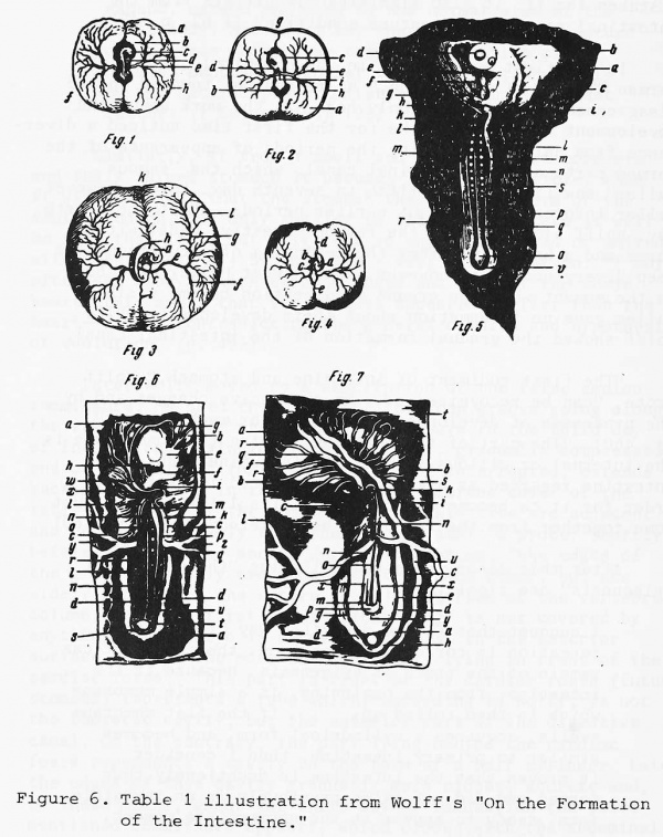
From the subsequent text it is clear, without doubt, that Wolff still could not differentiate the true preliminary membranes (serosa and amnion) , formed from the extra-embryonic parts of the ectoderm and mesoderm, from the embryonic layers, from which the embryo itself developed. According to his description, the condition of the egg about the middle of the third day of incubation is seen in Figure 6. A part of the yolk membrane, as Wolff called it, very rich in vessels, and the center of which is called the vascular area (area vasculosa) is where the embryo is found. This part is composed of two layers; the external layer is delicate, transparent and is deprived of vessels. It goes above the embryo and is loosely united with the internal layer so that during immersion in water it separates from it and comes to the surface. And the internal layer, together with the embryo, goes to the bottom. As a result, the embryo, which is still not surrounded by amnion, appears open'. The thin transparent film, easily separated from the embryo, which Wolff called the external layer, is nothing other than the yolk membrane, and the internal layer represents the blastodisc, actually composed of all three embryonic layers.
To the end of the third day and slightly later, according to Wolff's description, the vascular area also is composed of two layers: upper — thin — and lower — thicker and softer. But at this time they are so closely connected that they appear as one membrane. Near the embryo there is a transparent region; in its range both of these membranes begin to separate and the embryo becomes distinct. It appears between them so that the upper membrane passes above the embryo, and the lower passes under it.
Interpreting the description from the context of present knowledge about chick development, it can be suggested that the picture of separation of the two "membranes" corresponds to the formation of the exocoelom, i.e. the extraembryonic space, limited by the parietal and visceral layers of the mesoderm, so that Wolff's "upper membrane" is probably the ectoderm together with the parietal layer of the mesoderm, and "the lower membrane" is the visceral layer of the mesoderm together with the endoderm.
Wolff described the subsequent fate of the lower layer of the vascular area (Figure 7) as follows:
This layer goes from all sides of the vascular area in the direction of the embryo ... at a higher level , than the embryo and amnion . . . , as if it wants to pass through the amnion lying below with the embryo in it and to cover it. Only a small space remains in the middle of the upper surface of the amnion which is not covered with the internal layer ... After that, when the internal layer reaches this place, it is immediately turned in an acute angle, directed downwards, and lies around the embryo with the thin amnion surrounding it. After this, on the lower surface of the vascular area around the embryo, or more exactly around the amnion covering it, a vesicle must be formed ....
Wolff noticed that this vesicle can be seen distinctly, although it is not expressed identically in all eggs, and he considered that the other investigators did not notice it, because, in the first stages of incubation, they never examined the lower surface of the embryo lying on the yolk. In addition, he did not see this formation himself up to 1764, although he had investigated the developing eggs during the five previous years.
"All," Wolff continued, "who did not see the true amnion during its first appearance, or at least in the third or fourth day of incubation, assumed that this vesicle is the amnion, because the described vesicle is swollen and distended with fluid. It seems directly adjacent to the embryo, while the amnion itself, due to its extreme thinness and transparency, may remain unnoticed. Therefore, I called this formation the false amnion, although it does not properly possess any similarity with the true amnion. "4
It is not easy to translate into the language of recent embryology his description. Here, apparently, the descriptions of different formations are combined: on one hand Wolff dealt with the trunk folds, the apex of which was submerged inside the embryo. Finally this apex separates from the yolk sac. On the other hand, he saw a deepening of the endoderm, submerged from below in the embryo. All of this composed a picture of what Wolff described as the false amnion. 5
Cited places in § 36 and 37 under title "Vesicle, appearing in the lower surface of the transparent zone, or false amnion" (pp. 434 - 437) .
Nearly the same, K. M. Baer evaluated the idea of the Wolffian term as "false amnion," when he cited the work of Wolff in his classical work, HISTORY OF ANIMAL DEVELOPMENT (UBER ENTWICKELUNGSGESCHICHTE DER THIERE. BEOBACHTUNG UND REFLEXION, v. I, part I, 5 c)
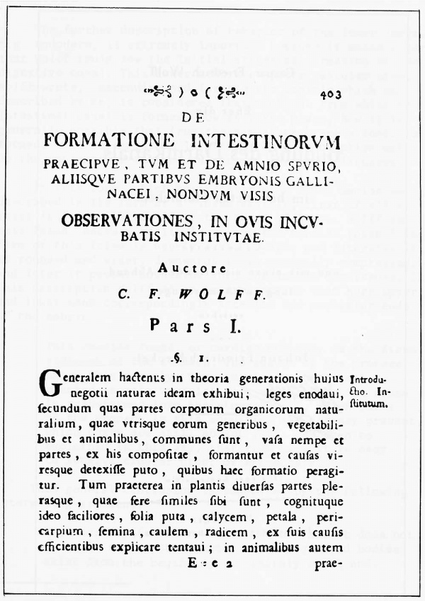
DF FORMATIONE INTESTINORVM PRAECIPVE , TVM ET DE AMNIO 5PVRIO, ALI1SQVE PART1BVS EMBRYONIS GALLl" NACEI , NONDVM VISIS OBSERVATIONES , IN OV1S EMCV BAT1S INST1TVTAE.
A u c to r c C. F. W L F F.
P a r s I.
i. Generalem haclcnus in tbeoria generationis huius Introdunegotii naturae ideam exhibui; leges enodaui, & i0 - In " fecundum quas partes corporum organicorum naturalium, quae vtrisque eorum genenbus , vegetabilibus et animalibus , communes funt , vafa nempe et partes , ex his compofitae , formantur et caufas vircsque detexifle puto , quibus haec formatio peragitur. Turn praeterea in plantis diucrfas partes plerasque , quae fere fimiles fibt funt , cognituque ideo faciliores , folia puta , calycem , petala , pericarpium , femina , caulem , radicem , ex fuis caufis efficientibus explicare tentaui ; in animalibus autem
E ; e a prae
Figure 4. The first page of Wolff's work "On the Formation of the Intestine."
Caspar Fnedrich Wolff iiber die Bildung des Darmkanals im bebruteten Huhnchen. Uebitietn uod mit einer einleitenden Abhand. lung und Anmerkungen verse liea
Figure 5. The title page of Meckel's translation of Wolff's work. Copy belonging to the first president of the Moscow Society of naturalists G. I. Fisher; kept in the library of the Society.
The further description of behavior of the lower layer, i.e. endoderm, is extremely important because it makes clear that Wolff truly saw the initial stages of formation of the digestive canal. This lower membrane of the vascular area, Wolff wrote, "ascending and forming the vesicle which was described by me, is considered that membrane from which the intestinal canal is formed. How it takes place, how it is generally possible that from the simple membrane a canal is formed, will be explained later on." This description makes up the contents of the second part of the work discussed.
In this zone, which Wolff called the false amnion and described in the form of a closed vesicle, first of all a fossa is formed, situated at the level of heart. Wolff called this fossa, because of its location, the cardiac fossa. 6 The form of this fossa is nearly oval. Upwards and laterally it is rounded and wider, downwards it is gradually compressed, and later it passes downwards into the amnion commissure. To this description Wolff added an explanation that here upper and lower mean correspondingly anterior and posterior ends of the embryo.
This cardiac fossa, or cardiac opening, is the first rudiment of the stomach. The result of the process shows this exactly. The vesicle is subsequently changed in this way: its part which forms the fossa is transformed into the stomach . . . Which parts of the stomach in this condition are already present, which parts are still absent . . . can then be explained in this way, that from here it is easy to receive clearer proof of epigenesis .
This general conclusion is supported by the following interesting argument.
From here it is clear without doubt that it does not occur in nature that parts of the organized bodies exist from the beginning, infinitely small and invisible but perfect and complete, and not thus, as if they were created instantly by the Most High Creator (22), and that finally, under the influence of accidental causes, as if stimulated, they begin to expand, extend and grow to the normal size of the adult body. Not so. I say that what I saw in nature occurs, but sooner, so that the formation of natural bodies in general is pre-established only by one natural power, existing in animal or plant matter. (5 56, p. 453) (23)
6. Meckel in his translation called it "gastric fossa" which maybe represents its role in development, but all this also may be unexactness of translation.
"Similarly, if from a small and invisible but complete and fully formed stomach it becomes large and visible, then it will be clear that the stomach then has the form of the adult stomach, but only smaller in size. In any case it will be seen that it is not half, not in any case opened or united with other parts" (§ 57, pp. 454 - 455). And later on: "I more often saw embryos with cardiac fossa and without the whole heart; this means that the stomach appeared earlier than the heart — a fact contradicting the general opinion and hypothesis of evolution" (p. 455).
Later Wolff moved to a description of the false amnion commissure. He used this name for the deep groove going along the ventral side of the embryo; it appears in the third day of incubation, beginning from the lower, gradually compressed end of the cardiac fossa and, not being interrupted, extends backwards, ending in the edge of the membrane cover of the tail. The edges of the commissures at this time are adjacent and united so closely that they do not admit a probe. Shortly before this, in the second day of incubation, "the edges of the groove not only are separated from each other, but are widely separated. The entire anterior surface of the vertebral column, which at first seems astonishing, is not covered by anything. The embryo is uncovered totally in the anterior surface, except the most anterior part, lying in front of the cardiac fossa." This part in front of the cardiac fossa (future stomach) represents a tube which, according to Wolff, is not the thoracic cavity, but the anterior part of the digestive canal. On the contrary, the part lying behind the cardiac fossa represents a cavity in the form of a half cylinder. Later, the edges of this cavity gradually move closer, accrete and, by these means, about the third day of incubation the above mentioned commissure appears, which closes with the abdominal side of the new-formed intestinal canal. "It must be noticed," IVolff wrote later, "that the intestinal canal, even when it is completely formed is so similar to the intestine that it may be mistaken for it; it also significantly differs from the intestinal canal in the mature condition" ($ 62, p. 450).
In his first works, even in the polemical part of the German "Theory of Generation," Wolff had not touched on the disagreement with Haller. Only here, in the work about the development of intestine, he for the first time noticed a divergence from Haller concerning the periods of appearance of the formed parts of the intestinal canal, which the "famous Haller" saw first in the fifth to seventh day. Later, however, Haller informed IVolff about earlier periods — about the fourth day. Wolff also observed the formed digestive canal within three and a half days after the beginning of incubation. The deep divergence lies, however, not in what Haller said but in the moment when the organs of digestion become visible. Haller gave no information about their development, while Wolff showed the gradual formation of the intestinal canal.
"The first rudiment of intestine and stomach," Wolff wrote, "can be recognized .... by successive changes and by the processes of development, which appear much earlier" (p. 460). "The part of the membrane forming the commissure is the internal or villous skin of the intestine. Thus, the intestine regarded as a whole is the opened intestine. In order for it to become a whole canal, its lateral parts must come together from the front and be accreted" (§ 63, p. 460).
After this description the following "thoughts about epigenesis" are significant.
I suggest that if the described way of intestine formation is correctly understood, then no doubt can remain of the truth of epigenesis. Because if the intestine, from the beginning, is a simple membrane which is then rolled up; . . .if the flat membrane swells, acquires a cylindrical form, and becomes similar to primary intestine, then I consider it proven that the intestine is doubtlessly thus formed and did not exist previously in an invisible form, ready to appear at the appropriate moment, (pp. 460 - 461)
Figure 6. Table 1 illustration from Wolff's "On the Formation of the Intestine . "
1 . Vascular area (area vasculosa) from upper surface after three days of incubation; a - vascular area; b - transparent place (area pelludica) ; c - embryo; d - heart; e - lateral yolk veins; f - descending vein; k - boundary vein.
2. Lower surface of the vascular area and on it the vesicle or false amnion; a - lower surface of the vascular area; b - transparent place; c - false amnion and on it (from c to d) cephalic branch; di - middle part of it; i to b - caudal membrane; d - cardiac fossa, from d to i, commissure; i - lower fossa; e - lateral veins; f - descending vein; g - ascending vein; h - boundary vein.
3. Upper surface of the vascular area in living embryo of three and a half days of incubation; a - embryo; b - fossa, which it forms in yolk; c - origin of yolk veins; d - their lower branches; f - middle branches; g - branching of the upper branches; h - ascending vein; k - its branches; i - descending vein; 1 - terminal vein.
4. Vascular area in living condition; a - chamber of heart; from above, bulb of aorta, from downwards, auricle of the heart; b - aorta; c - hollow vein; d - ascending vein; e - descending vein.
5. Transparent place in the embryo from lower surface, under microscope, 54 hours of incubation; a - transparent place; b - cephalic branch; c - part of the embryo above the cardiac fossa; d - anterior part of the head;
e - heart; f - first rudiment of the true amnion; g - ascending vein; h - origin of cephalic branch; i - swollen edge of the ventricle opening; k - opening of the stomach still is not closed, but has narrowed; 1 - abdominal folds; m - intestinal folds; n - rudiments of vertebrae; o - end of vertebrae; p - the beginning of formation of amnion opening; q - spinal cord; r - marked lateral parts of the vesicle; s - traces of retiform, derived from red blood, vessels; v- -first rudiment of the caudal membrane.
6. Part of lower surface of the vascular area with the transparent place and embryo, after two days of incubation.
a - the transparent place; b - cephalic branch; c - upper, swollen region of the lateral part of the vesicle; d - lower, still not swollen part; e - part of the embryo over the cardiac fossa; f - heart; g - occiput; h - anterior part of the head; i - edge of the cardiac fossa; k - left abdominal fold; 1 - right lower abdominal fold; m - internal intestinal fold; n - right intestinal fold; o- spinal cord; p- cavity of the intestinal canal; q, r - lateral deepenings, rudiments of cavity of the body; from s to t - rudiment of caudal membrane; s - united abdominal folds; t- union of the intestinal folds, origin of rectum; u - slightly bending end of the spinal chord; w- ascending vein; x - its branches; y- right lateral vein; z - left lateral vein.
7. Part of the vascular area, containing the embryo, covered by the false amnion, after 60 hours of incubation, a - immovable false amnion; b - cephalic branch; c - lateral parts of the vesicle; d - caudal membrane; e, f, i, g - embryo, translucent through the vesicle; f - occiput; g - protuberance of right leg; h - rudiment of tailbone; i - protuberance of left wing; k - cardiac fossa; 1 - commissure; m - lower fossa; n - fold of commissure; o - opening between abdominal and intestinal folds; p- fold of lower fossa; q - ascending vein; r - its branches; s- - vein associated with the ascending; t - its branches; u - descending vein; x - abdominal fold; y - area formed by the union of trunk folds.
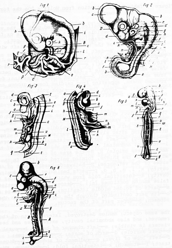
Figure 7. Table II illustration from Wolff's "On the Formation of the Intestine . "
1. Embryo of 4 days incubation; r - excised part of the vascular area on which the vesicle lies; a, b, c - vesicle or false amnion; g, h, i, 1- - translucent embryo; h - occiput;
g - anterior part of the head; i - vertebra; 1 - left wing; k - left auricle; B - left lateral part of thorax; C - fold of abdominal opening and true amnion (Table I , Fig. 2, ii, Fig. 7, xx) ; e - intestinal fold and fold of false amnion (see Table I, Fig. 7, n) ; m - opening between true and false amnion; d - part of middle intestine; f - rounded fossa, appearing from cardiac fossa, groove and lower fossa; N - lower fossa; p - right yolk vein; o - artery of that side; n - left yolk artery; q- -umbilical vesicle (allantois) .
2. The same object with extracted false amnion; a - part of upper layer of vascular area; b- -true amnion; c - occiput; d - anterior part of the head; D- middle protuberance, disappearing after six days; e- -vertebra; f - left wing;
F - left leg; g - left auricle; h- - lateral part of thorax; i - folds of abdominal opening from which membrane of true amnion takes origin; H, H- -the area of the abdomen, adjacent to the thighs, forming paired cavities in the sides of the vertebrae; E- - backwards-bending part of this cavity; k, 1, m, n, o, p- parts beginning from abdominal cavity; k - intestinal fold from which the membrane of the false amnion continues (n) ; 1 - deeper fold of intestine; m - parts of folded middle intestine; n - part of false amnion; o - its part continuing through the cardiac fossa to the stomach; p- upper part of the rectum.
3. The same object; amnion destroyed, lateral parts of thorax removed; and also abdominal cavity, extremities and head; viscera is visible; a - left chamber of heart; b - aural canal; c - left auricle; d - arch of aorta; v- - venous sinus; f - left lobe of liver; g - rudiment of abdomen; h - digestive tract; i - stomach; k - intestinal fold; 1 - duodenum; MM - middle intestine; n - part of membrane of false amnion; o - part of this membrane covering the intestinal canal; p- upper funnel-shaped part of the rectum; q - lower part of rectum, which, like the stomach, also already has cylindrical form, while lying between them a part of intestine opened; r - mesentery - continuation of false amnion and intestinal membranes; s - left kidney; t - part of vertebrae; u - left yolk artery.
4. The same preparation from the right side; a- - left chamber of the heart; b - its right chamber; c - aorta; d - arch of aorta; e - rudiment of left lung; f - right lobe of liver;
g - right kidney; h - right yolk artery; i - right intestinal membrane, from which middle intestine is formed; k - rectum; 1- mesentery; m - right membrane of false amnion; n - right yolk vein; N - trunk of vein; o - vertebrae.
5. Embryo represented in Table 1, Fig. 6, liberated from all membrane; a - anterior part of the head; b- - occiput; c, d - occipital region; d, e - region of thorax; e-z- the rest of the vertebrae; g - left part of the lower jaw; f - -process (first rib) ; h - left auricle; i - aural canal; k - left chamber of heart; 1 - aorta; m - membrane represented in Fig. 3, g; n - part of false amnion from which the cardiac fossa and stomach are formed; o, p, q, s, t, z - first rudiment of abdomen in the form of bending membrane; o - upper part of this membrane; p, s- edge of abdominal fold; q - its upper left, strongly curved part; t - its edge in the place where it becomes wider; v - right lateral abdominal membrane; u - left membrane; s, r, z - intestinal canal (fistula intestinalis) - first rudiment of mesentery with still divided membranes; s - right mesenterical membrane with right kidney; r - the same also - left; w- - opening between membranes of mesentery, in which uncovered vertebra is seen; x - vertebrae; y - rudiment of tailbone; z - rudiment of the pelvis .
6 . Embryo represented in Table I , Fig . 7 ; a - anterior part of the head; b - posterior part of the head; c - protuberance (see Fig. 2, D) ; d - thoracic part of the vertebrae; e - occipital region; f - rudiments of ribs; g - rudiment of wing; h - rudiment of leg; i - hip region; k - region of tailbone; 1 - rudiment of pelvic cavity; m - rudiment of true amnion; n - part of thorax; o - left auricle; q- - middle chamber; r a bdomen ; s - stomach; t - ascending vein; u - part of cephalic branch; v - edge of the stomach (see Table I, Fig. 1 , k) -, w - intestinal fold; x - part of false amnion; y - completely formed mesentery; z - upper, funnel-shaped part of the rectum; A - left, kidney, later on separated from the mesentery; B - yolk artery.
Wolff illustrated the principle of gradual establishment of parts in the process of development with many examples. He referred to the description and drawings of Rezel, according to which in the tadpoles of frogs^ there are no legs at the beginning. The extremities are developed later. He said later that, according to Reaumer, the accidentally destroyed chelae of crayfish grow again and that Trembley saw simple polyps which later became branched and complicated (his report, apparently, is about the regeneration of antennae and about the budding of hydra) .
Describing the gradual formation of the intestinal canal from the rolled-up and accreted edges of the membrane, Wolff noticed, though obscurely, the similar formation of the central nervous system. In fact, his analogy confirms the general principle of gradual development for the commissure of the false amnion, for the primary intestine, for the brain and spinal cord, and even for the embryo as a whole. Wolff's discovery of the common manner of development led to his assumption of the transformation of that which is flat by means of closure of its edges, into a hollow cylindrical body. Apparently Wolff could not observe directly the process of rolling up of the medullary membrane in a canal. In any case, the propagation of the described principle of development on the central nervous system, even if an assumption at the time, was an excellent discovery which Baer made sixty years later.
In addition Wolff turned attention to the successive formation of the different systems of organs, the first of which, in his opinion, is the central nervous system. Then, he supposed, the muscles are isolated. The third to become clear is the circulatory system, and, finally, the digestive system (p. 472) .
From the third part of the reviewed work we must consider only the general conclusions, which partly repeat those above, but which deserve further mention because here Wolff brought a final summation of the theory of epigenesis as opposed to the idea of preexistence of the parts of the embryo. Thus, he wrote :
We see that many parts of the body, for example the thorax, at a certain moment of time not only do not exist completely, but also cannot exist at that time. We conclude that it did not exist not because we do not observe it, but because we see in this place, where the thorax must appear, the appearance of true amnion; from here we conclude that the thorax, which is not revealed, cannot exist and as a matter of fact does not exist.
These arguments of Wolff also concern development of the pelvis and the digestive tract. "I consider," he concluded, "that this is the most important evidence in favor of epigenesis."
From here it can be concluded that the parts of the body do not always exist, but are formed gradually; during this it is not important by what means formation is accomplished; I do not say that the parts are formed by the accumulation of particles , or by any kind of fermentation, by means of mechanical causes or by powers of soul ; I only say that the parts are truly formed .... Instead of the center of the intestine, i.e. the entire intestinal tract from duodenum to rectum, there are two plates with rolled up anterior edges, and in the rest plates divided are falling behind each other .... I ask, therefore, are these plates the formed intestine? No one, of course, will confirm this . Thus , I conclude that the complete and formed parts do not always exist, but are formed in a determined period after conception. ($ 155)
This necessarily brief and selective statement of Wolff's ideas gives a clear view of the means of exact investigation of embryonic development by which he finally consolidated and confirmed the truth of epigenetic opinions and the fruitlessness of the preformation idea.
Wolff was insufficiently evaluated by contemporaries. Forty years later the majority of them had no idea about the existence of his main work. Meckel's merit is not only that by publishing his translation he made Wolff's work general property, but also that he helped restore Wolff's priority and credited him with "contributing to the history of development in general and development of the intestine in particular."
The evaluation of Wolff's work was given by Meckel in the following statements:
This exactness of observation, this step-by-step tracing of organs from their first appearance to their final development, only this could lead to creation of a true history of embryonic development. The author did not follow preconceived opinions, did not report more than he saw, and did not promote only probable propositions as law. The work is considered a model in all respects, so that I do not think that I can be reproached for translating the work of 1768. Knowing that it remained almost completely unknown, and also that it was found in a journal, available to only a few readers, completely eliminates my indecision
"This work until this time remained unknown to the physiologists," Meckel continued, "as attested to by the fact that Oken, who published in 1806 an article about intestine formation, did not possess any knowledge of Wolff's work, as he did not mention it anywhere" (Introductory article, p. 5). In many following pages Meckel compared Oken's results with Wolff's data and showed that Wolff had established much long before Oken.
Wolff's discoveries were much ahead of his time; he remained misunderstood by the majority of his contemporaries. About forty years after Wolff's death, his great successor in the Petersburg Academy, K. M. Baer, said the following: "Not only superiority of work assured its success .... Academician Wolff, after untiring work, discovered tb? law of organic transformation; but at that time the time lad not yet come to investigate this law, and science disregarded him (24) . After half a century, others succeeded with little reinforcement in obtaining laurels themselves in this same field, noticed his existence, and exalted the memory of Wolff." 7 As is well known, Baer praised Wolff's work on the intestine. °
(Footnotes on next page)
The American zoologist Wheeler, in the past century, wrote about the significance of Wolff's embryo logical works in the following poetic words: "The Siegfried destined to overcome this monstrous theory of emboitement, a theory not only false in itself, but one jealously guarding the problem of development, and preventing all access to it, as the dragon guarded the treasure of the Niebelungen, was CASPAR FRIEDRICH WOLFF. "9
The contemporary historian of embryology J. Needham also highly evaluated the significance of Wolff's work for refuting the idea of preformation of the embryo. Needham, repeating the expression of Claude Bernard, -10 considered that "this was a fatal blow for the theory of preformation.^
7 . Opinion about the development of science , speech read October 24, 1835 at the public meeting of the Academy of Science by Baer. JOURN . MIN. NAR. PROSV., May 1836, pp. 190 - 245 (citation p. 218).
8. K. E. V. Baer, UBER ENTWICKELUNGSGESCHICHTE DER THIERE. BEOBACHTUNG UND REFLEXION, 1837, v. II, part 3, 7 (footnote) .
9. W. M. Wheeler, "Caspar Friedrich Wolff and Theoria generationis . " BIOL. LECTURES, Mar. biol . lab. Wood's Hole, 1898 - 99, p. 271.
10. Claude Bernard, LECONS SUR LES PHENOtyENES I>E LA VIE COMMUNE AUX ANIMAUX ET AUX VEGETAUX. (Paris: J. B. Bailliere et fils, 1878 - 1879), p. 316.
11. J. Needham, A HISTORY OF EMBRYOLOGY, p. 258.
Cite this page: Hill, M.A. (2024, April 26) Embryology Book - Russian Embryology (1750 - 1850) 5. Retrieved from https://embryology.med.unsw.edu.au/embryology/index.php/Book_-_Russian_Embryology_(1750_-_1850)_5
- © Dr Mark Hill 2024, UNSW Embryology ISBN: 978 0 7334 2609 4 - UNSW CRICOS Provider Code No. 00098G


