Bone Development: Difference between revisions
No edit summary |
No edit summary |
||
| Line 90: | Line 90: | ||
* '''Endocrinology: An Integrated Approach''' by Nussey, S.S. and Whitehead, S.A. [http://www.ncbi.nlm.nih.gov:80/books/bv.fcgi?db=Books&rid=endocrin.box.936 Endocrinology: Definition and causes of osteoporosis] | * '''Endocrinology: An Integrated Approach''' by Nussey, S.S. and Whitehead, S.A. [http://www.ncbi.nlm.nih.gov:80/books/bv.fcgi?db=Books&rid=endocrin.box.936 Endocrinology: Definition and causes of osteoporosis] | ||
* '''Developmental Biology''' 6th ed. by Gilbert, Scott F. [http://www.ncbi.nlm.nih.gov:80/books/bv.fcgi?db=Books&rid=dbio.section.4388#4391 Aging: The Biology of Senescence] | * '''Developmental Biology''' 6th ed. by Gilbert, Scott F. [http://www.ncbi.nlm.nih.gov:80/books/bv.fcgi?db=Books&rid=dbio.section.4388#4391 Aging: The Biology of Senescence] | ||
[[Category:Musculoskeletal]] [[Category:Bone]] | |||
Revision as of 13:41, 21 September 2009
Introduction
Macroscopic and microscopic bone structure in the adult and during development.
For more development background see Science Lecture - Musculoskeletal Development
Textbook
Histology and Cell Biology: An Introduction to Pathology, A.L. Kierszenbaum, 2002 - Connective Tissue, Chapter 4 pp118-129; Osteogenesis, Chapter 5 pp131-145
Slides
UNSW Virtual Slidebox Virtual Slidebox Phase 1
- Compact Bone - Adult Bone , Adult (Ground, TS) Human Alizarin Red | Bone (Ground, LS) Human Alizarin Red
- Compact Bone Rib Decalcified rib, bone marrow
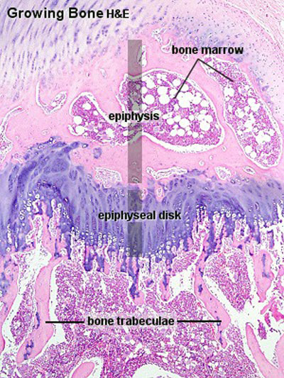
- Endochondral ossification Developing bone | Bone, Developing (LS, Femur) Cat H&E
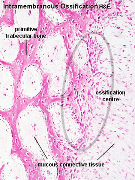
- Intramembranous ossification Head (Neonatal) Rat H& Van Gieson
Haversian Systems
- also called osteons
- Volkmann's canals
Lamellae
- concentric - surrounding each Haversian System
- interstitial - bony plates that fill in between the haversian systems.
- circumferential - layers of bone that underlie the periosteum and endosteum
Cells
- osteocytes and canaliculi
Bone Structure
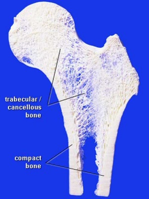
Compact bone
- no spaces or hollows in the bone matrix visible to the eye.
- forms the thick-walled tube of the shaft (or diaphysis) of long bones, which surrounds the marrow cavity (or medullary cavity). A thin layer of compact bone also covers the epiphyses of long bones.
Trabecular bone
- (cancellous or spongy bone) consists of delicate bars (spicules) and sheets of bone, trabeculae
- branch and intersect to form a sponge-like network
- ends of long bones (or epiphyses) consist mainly of trabecular bone.
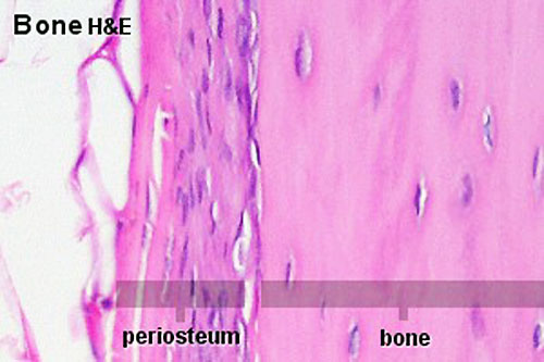
Periosteum
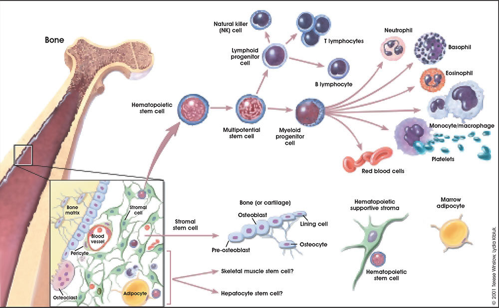
Bone Cells
Osteocytes
- mature bone-forming cells embedded in bone matrix
- derive from osteogenic stem cells forming Osteoblasts
- line surface of bone, secrete organic matrix of bone (osteoid), converted into osteocytes when become embedded in matrix (which calcifies soon after deposition) (Google- osteoblast images)
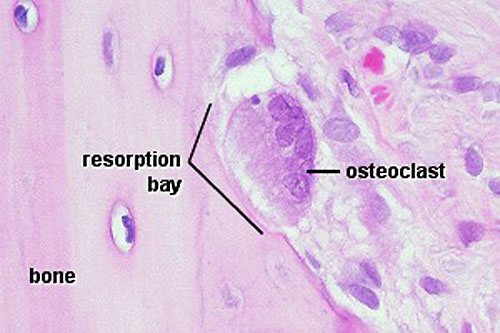
Osteoclasts
- bone-resorbing multinucleated macrophage-like cells
- origin- fusion of monocytes or macrophages, Blood macrophage precursor, Attach to bone matrix
- seal a small segment of extracellular space (between plasma membrane and bone surface), HCl secreted into this space by osteoclasts dissolves calcium phosphate crystals (give bone rigidity and strength) (Google- osteoclast images)
- do not mistake for megakaryocytes, found in bone marrow not associated with bone matrix.
- megakaryocytes are also multi-niucleated and form platelets
Bone Matrix
The bone matrix has 2 major components. Organic portion composed of mainly collagen Type 1 (about 95%) and amorphous ground substance. Inorganic portion (50% dry weight of the matrix) composed of hydroxyapatite crystals, calcium, phosphorus, bicarbonate, nitrate, Mg, K, Na. (Google- bone matrix images)
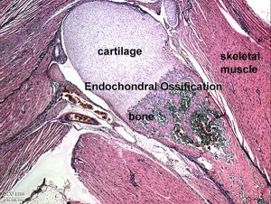
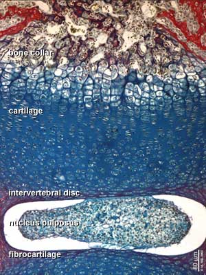
Endochondral ossification
Endochondral ossification slides Developing bone | Bone, Developing (LS, Femur) Cat H&E
Intramembranous ossification
Links
- Original class notes
- UNSW Virtual Slidebox Virtual Slidebox Phase 1
- Virtual Slidebox of Histology (USA) Skeletal system
- Lecture - Musculoskeletal Development
Other Textbooks
- Anatomy of the Human Body (H. Gray, 1918.) historical anatomy text Osteology
- Molecular Biology of the Cell Bone Is Continually Remodeled by the Cells Within ItImage: Figure 22-52. Deposition of bone matrix by osteoblasts.Image: Figure 22-56. The development of a long bone.
- Molecular Cell Biology Mutations in Collagen Reveal Aspects of Its Structure and Biosynthesis
- The Cell- A Molecular Approach Steroid Hormones and the Steroid Receptor Superfamily
- Clinical Methods: The History, Physical, and Laboratory Examinations 100. Alkaline Phosphatase and Gamma Glutamyltransferase
- Endocrinology: An Integrated Approach by Nussey, S.S. and Whitehead, S.A. Endocrinology: Definition and causes of osteoporosis
- Developmental Biology 6th ed. by Gilbert, Scott F. Aging: The Biology of Senescence
